. 2002 Dec 2;196(11):1515-21.
doi: 10.1084/jem.20011794.

Adhesion molecule-dependent mechanisms regulate the rate of macrophage clearance during the resolution of peritoneal inflammation

Affiliations

Adhesion molecule-dependent mechanisms regulate the rate of macrophage clearance during the resolution of peritoneal inflammation

Geoffrey John Bellingan et al. J Exp Med. .

Abstract

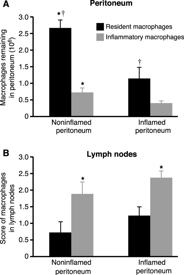
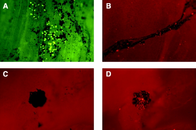
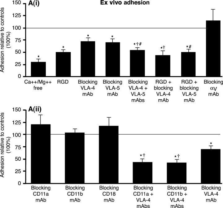
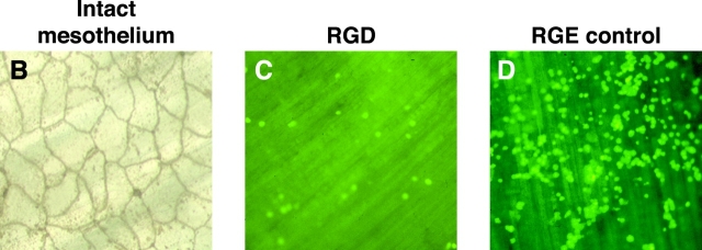
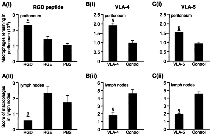
Macrophage clearance is essential for the resolution of inflammation. Much is known about how monocytes enter the inflammatory site but little is known about how resultant macro-phages are cleared. We have previously demonstrated that macrophage clearance from resolving peritonitis occurs by emigration into draining lymphatics rather than local apoptosis. We now examine mechanisms for this process, in particular by evaluating the hypothesis that modulation of adhesion interactions between macrophages and cells lining the lymphatics regulates the rate of macrophage clearance. We demonstrate in vivo that macrophages adhere specifically to mesothelium overlying draining lymphatics and that their emigration rate is regulated by the state of macrophage activation. We observed that macrophage-mesothelial adhesion is Arg-Gly-Asp (RGD) sensitive and partially mediated by very late antigen (VLA)-4 and VLA-5 but not alpha(v) or beta(2) integrins. Moreover, macrophage clearance into lymphatics can be blocked in vivo by RGD peptides and VLA-4 and VLA-5 but not beta(2) blocking antibodies. This is the first evidence that macrophage emigration from the inflamed site is controlled and demonstrates that this is exerted through specific adhesion molecule regulation of macrophage-mesothelial interactions. It highlights the importance of adhesion molecules governing entry of cells into the lymphatic circulation, thus opening a new avenue for manipulating the resolution of inflammation.


Figures

Figure 1.
Figure 1.
Macrophage emigration is regulated. A total of 12.5 × 10⁶ fluorescent green–labeled resident and red-labeled inflammatory macrophages were instilled into the peritoneal cavity of mice in the absence of peritonitis (noninflamed peritoneum) or mice with resolving peritonitis (inflamed peritoneum). (A) The number of resident (black bars) and inflammatory (gray bars) macrophages recovered from the peritoneal cavity after 3 d is shown. † significantly greater resident than inflammatory macrophage recovery (P < 0.05, n = 8/group). *, significantly greater recovery from noninflamed than inflamed peritoneum (P < 0.05). (B) Fluorescent-labeled macrophages in draining parathymic lymph nodes from the same mice. *, significantly more inflammatory than resident macrophages (P < 0.05, n = 8/group).
Figure 2.
Figure 2.
Macrophage–mesothelial adhesion is localized to regions overlying the draining lymphatics. (A) Ex vivo preparation of mesothelial surface of diaphragm with lymphatics localized by India ink. Adherence of inflammatory macrophages localizes to areas of mesothelium stained with India ink whereas virtually no macrophages adhere to India ink–free areas. Similar colocalization is seen when adhesion occurs in vivo. (B) Macrophages emigrating into draining lymphatics within 1 h of instillation. (C) India ink labeling of a milky spot and (D) the same section under fluorescent microscopy where fluorescent macrophages are seen to adhere (×80).
Figure 3.
Figure 3.
β1 integrins mediate macrophage–mesothelial adhesion. (A) (i) Macrophage–mesothelial adhesion ex vivo is significantly reduced (P < 0.05) by the following: Ca2+/Mg2+-free media (control: Ca2+/Mg2+-containing media, n = 5), 0.5 mg/ml RGD in standard media (control: RGE, n = 5), blocking: VLA-4 (PS/2 10 μg/ml, n = 6), VLA-5 (5H10–27 10 μg/ml, n = 9), VLA-4 + VLA-5 (n = 7), VLA-4 + RGD (n = 9), and VLA-5 + RGD (n = 8), but not by blocking αv (H9.2B8, 10 μg/ml, n = 6). Controls (isotype monoclonals, F4/80 and/or RGE) represent 100% adhesion. Adhesion significantly reduced compared to the following: controls (*), blocking VLA-4 (†), and blocking VLA-5 (#). (ii) Blocking β2 integrins: CD11a (M17/4, n = 8), CD11b (M1/70, 5C6 both n = 6), and CD18 (GAME-46, 2E6 both n = 9) do not inhibit adhesion, however blocking both β1 and β2 integrins, VLA-4 + CD11a (n = 6) and VLA-4 + CD11b (M1/70, n = 6), inhibits adhesion significantly more than blocking β1 integrins alone (PS/2, n = 6). Adhesion was significantly reduced (P < 0.05) compared to the following: controls (*) and blocking VLA-4 (†). (B) En face silver staining demonstrating the integrity of mesothelial surface after 6 h is shown (×300). (C and D) Adhesion of fluorescent-labeled macrophages to mesothelium ex vivo in the presence of RGD or RGE, which shows the ease of visual quantification of assay (×100).
Figure 3.
Figure 3.
β1 integrins mediate macrophage–mesothelial adhesion. (A) (i) Macrophage–mesothelial adhesion ex vivo is significantly reduced (P < 0.05) by the following: Ca2+/Mg2+-free media (control: Ca2+/Mg2+-containing media, n = 5), 0.5 mg/ml RGD in standard media (control: RGE, n = 5), blocking: VLA-4 (PS/2 10 μg/ml, n = 6), VLA-5 (5H10–27 10 μg/ml, n = 9), VLA-4 + VLA-5 (n = 7), VLA-4 + RGD (n = 9), and VLA-5 + RGD (n = 8), but not by blocking αv (H9.2B8, 10 μg/ml, n = 6). Controls (isotype monoclonals, F4/80 and/or RGE) represent 100% adhesion. Adhesion significantly reduced compared to the following: controls (*), blocking VLA-4 (†), and blocking VLA-5 (#). (ii) Blocking β2 integrins: CD11a (M17/4, n = 8), CD11b (M1/70, 5C6 both n = 6), and CD18 (GAME-46, 2E6 both n = 9) do not inhibit adhesion, however blocking both β1 and β2 integrins, VLA-4 + CD11a (n = 6) and VLA-4 + CD11b (M1/70, n = 6), inhibits adhesion significantly more than blocking β1 integrins alone (PS/2, n = 6). Adhesion was significantly reduced (P < 0.05) compared to the following: controls (*) and blocking VLA-4 (†). (B) En face silver staining demonstrating the integrity of mesothelial surface after 6 h is shown (×300). (C and D) Adhesion of fluorescent-labeled macrophages to mesothelium ex vivo in the presence of RGD or RGE, which shows the ease of visual quantification of assay (×100).
Figure 4.
Figure 4.
VLA-4 and VLA-5 regulate macrophage clearance from resolving peritonitis. (A) (i) After fluorescently labeling macrophages in vivo, 0.4 mg RGD or controls, RGE (0.4 mg) or PBS, were injected 2 and 26 h later and macrophage recovery from peritoneum was determined 24 h later. This was significantly greater for RGD than controls. *, P < 0.05. n = 13 RGD, 8 RGE, and 10 PBS. Controls were not different. (ii) RGD significantly reduced the number of macrophages reaching draining lymph nodes compared with RGE or PBS (§, P < 0.01). (B) (i) Blocking VLA-4 (PS/2) led to significantly more macrophages remaining in the peritoneum (§, P < 0.01; n = 10 VLA-4; 9 controls) and (ii) significantly less in the lymph nodes (§, P < 0.01) than isotype control. (C) (i) Blocking VLA-5 (5H10–27) led to significantly more macro-phages remaining in the peritoneum (§, P < 0.01; n = 10 VLA-5; 10 controls) and (ii) significantly less in the lymph nodes (§, P < 0.01) than isotype control.

References

    1. Haslett, C. 1999. Granulocyte apoptosis and its role in the resolution and control of lung inflammation. Am. J. Respir. Crit. Care Med. 160:S5–S11. - PubMed
    1. Bellingan, G.J., H. Caldwell, S.E. Howie, I. Dransfield, and C. Haslett. 1996. In vivo fate of the inflammatory macrophage during the resolution of inflammation: inflammatory macrophages do not die locally, but emigrate to the draining lymph nodes. J. Immunol. 157:2577–2585. - PubMed
    1. Van Furth, R. 1992. Development and distribution of mononuclear phagocytes. Inflammation: Basic Principles and Clinical Correlates. Second Edition. J.I. Gallin, I.M. Goldstein, and R. Snyderman, editors. Raven Press, New York. 325–341.
    1. Leibovich, S.J., and R. Ross. 1975. The role of the macrophage in wound repair. A study with hydrocortisone and anti-macrophage serum. Am. J. Pathol. 78:71–100. - PMC - PubMed
    1. Adams, D.O., and T.A. Hamilton. 1992. Macrophages as destructive cells in host defence. Inflammation: Basic Principles and Clinical Correlates. Second Edition. J.I. Gallin, I.M. Goldstein, and R. Snyderman, editors. Raven Press, New York. 637–643.

Publication types