

Site-directed recombination via bifunctional PNA-DNA conjugates
- PMID: 12461167
- PMCID: PMC139206
- DOI: 10.1073/pnas.262556899
Site-directed recombination via bifunctional PNA-DNA conjugates
Abstract
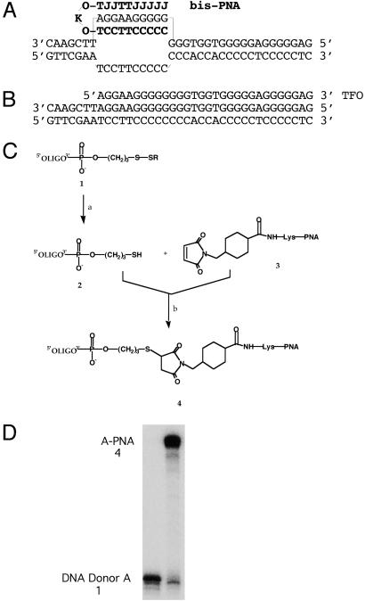
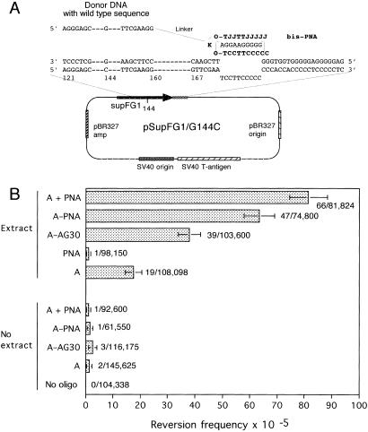
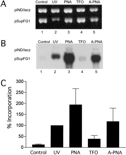
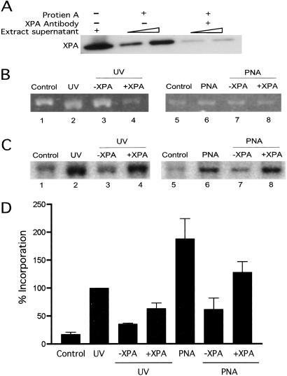
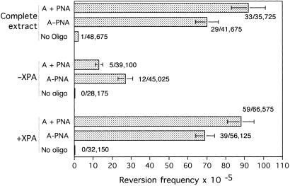
Site-specific DNA binding molecules offer the potential for genetic manipulation of mammalian cells. Peptide nucleic acids (PNAs) are a DNA mimic in which the purine and pyrimidine bases are attached to a polyamide backbone. PNAs bind with high affinity to single-stranded DNA via Watson-Crick base pairing and can form triple helices via Hoogsteen binding to DNAPNA duplexes. Dimeric bis-PNAs capable of both strand invasion and triplex formation can form clamp structures on target DNAs. As a strategy to promote site-directed recombination, a bis-PNA was coupled to a 40-nt donor DNA fragment homologous to an adjacent region in the target gene. The PNA-DNA conjugate was found to mediate site-directed recombination with a plasmid substrate in human cell-free extracts, resulting in correction of a mutation in a reporter gene at a frequency at least 60-fold above background. Induced site-specific recombination was also seen when the bis-PNA and the donor DNA were co-mixed without covalent linkage. In addition, the bis-PNA and the bis-PNA-DNA conjugate were found to induce DNA repair specifically in the target plasmid. Both the PNA-induced recombination and the PNA-induced repair were found to be dependent on the nucleotide excision repair factor, XPA (xeroderma pigmentosum complementation group A protein). These results suggest that the formation of a PNA clamp on duplex DNA creates a helical distortion that strongly provokes DNA repair and thereby sensitizes the target site to recombination. The ability to promote recombination in a site-directed manner using PNA-DNA conjugates may provide a useful strategy to achieve targeted correction of defective genes.
Figures
Similar articles
-
Site-specific gene modification by PNAs conjugated to psoralen.Biochemistry. 2006 Jan 10;45(1):314-23. doi: 10.1021/bi051379a. Biochemistry. 2006. PMID: 16388608
-
PNA-nitrogen mustard conjugates are effective suppressors of HER-2/neu and biological tools for recognition of PNA/DNA interactions.Bioconjug Chem. 2006 Jan-Feb;17(1):214-22. doi: 10.1021/bc0502964. Bioconjug Chem. 2006. PMID: 16417271
-
An experimental study of mechanism and specificity of peptide nucleic acid (PNA) binding to duplex DNA.J Mol Biol. 1999 Mar 12;286(5):1337-45. doi: 10.1006/jmbi.1998.2578. J Mol Biol. 1999. PMID: 10064701
-
Peptide nucleic acids (PNA) and PNA-DNA chimeras: from high binding affinity towards biological function.Biol Chem. 1998 Aug-Sep;379(8-9):1045-52. Biol Chem. 1998. PMID: 9792437 Review.
-
Triplex DNA structures.Annu Rev Biochem. 1995;64:65-95. doi: 10.1146/annurev.bi.64.070195.000433. Annu Rev Biochem. 1995. PMID: 7574496 Review.
Cited by
-
Targeted gene conversion induced by triplex-directed psoralen interstrand crosslinks in mammalian cells.Nucleic Acids Res. 2009 Oct;37(19):6378-88. doi: 10.1093/nar/gkp678. Epub 2009 Sep 2. Nucleic Acids Res. 2009. PMID: 19726585 Free PMC article.
-
Poly(Lactic-co-Glycolic Acid) Nanoparticle Delivery of Peptide Nucleic Acids In Vivo.Methods Mol Biol. 2020;2105:261-281. doi: 10.1007/978-1-0716-0243-0_17. Methods Mol Biol. 2020. PMID: 32088877 Free PMC article.
-
Peptide Nucleic Acids as a Tool for Site-Specific Gene Editing.Molecules. 2018 Mar 11;23(3):632. doi: 10.3390/molecules23030632. Molecules. 2018. PMID: 29534473 Free PMC article. Review.
-
Systemic in utero gene editing as a treatment for cystic fibrosis.Proc Natl Acad Sci U S A. 2025 Jun 17;122(24):e2418731122. doi: 10.1073/pnas.2418731122. Epub 2025 Jun 10. Proc Natl Acad Sci U S A. 2025. PMID: 40493185
-
Next generation triplex-forming PNAs for site-specific genome editing of the F508del CFTR mutation.J Cyst Fibros. 2025 Jan;24(1):142-148. doi: 10.1016/j.jcf.2024.07.009. Epub 2024 Aug 5. J Cyst Fibros. 2025. PMID: 39107154
References
Publication types
MeSH terms
Substances
Grants and funding
LinkOut - more resources
Full Text Sources
Other Literature Sources
Research Materials
Miscellaneous
