

A putative molecular-activation switch in the transmembrane domain of erbB2
- PMID: 12461170
- PMCID: PMC138543
- DOI: 10.1073/pnas.252640799
A putative molecular-activation switch in the transmembrane domain of erbB2
Abstract
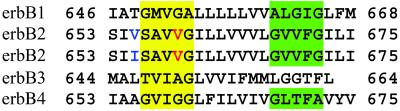
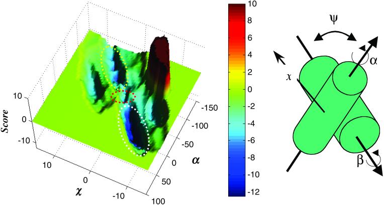
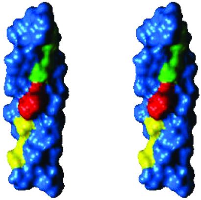
Overexpression of the receptor tyrosine kinase (RTK) erbB2 (also designated neu or HER2) was implicated in causing a variety of human cancers, including mammary and ovarian carcinomas. Ligand-induced receptor dimerization is critical for stimulation of the intrinsic protein tyrosine kinase (PTK) of RTKs. It was therefore proposed that PTK activity is stimulated as a result of the reorientation of the cytoplasmic domains within receptor dimers, leading to transautophosphorylation and stimulation of enzymatic activity. Here, we propose a molecular mechanism for rotation-coupled activation of the erbB2 receptor. Using a computational exploration of conformation space of the transmembrane (TM) segments of an erbB2 homodimer, we found two stable conformations of the TM domain. We suggest that these conformations correspond to the active and inactive states of erbB2, and that the receptor molecules may switch from one conformation to the other without crossing exceedingly unfavorable states. This model provides an explanation for the biochemical and oncogenic properties of erbB2, such as the effects of erbB2 overexpression on kinase activity and cell transformation. Furthermore, the opposing effects of the neu* activating oncogenic point mutation and the Val-655-->Ile single-nucleotide polymorphism shown to be linked to reduced risk of breast cancer are explained in terms of shifts in the equilibrium between the active and inactive states of erbB2 in vivo.
Figures
Similar articles
-
Dimer interface of transmembrane domains for neu/erbB-2 receptor dimerization and transforming activation: a model revealed by molecular dynamics simulations.J Biomol Struct Dyn. 2001 Aug;19(1):15-31. doi: 10.1080/07391102.2001.10506717. J Biomol Struct Dyn. 2001. PMID: 11565846
-
The ErbB/HER receptor protein-tyrosine kinases and cancer.Biochem Biophys Res Commun. 2004 Jun 18;319(1):1-11. doi: 10.1016/j.bbrc.2004.04.150. Biochem Biophys Res Commun. 2004. PMID: 15158434 Review.
-
Sequence-dependent oligomerization of the Neu transmembrane domain suggests inhibition of "conformational switching" by an oncogenic mutant.Biochemistry. 2010 Apr 6;49(13):2811-20. doi: 10.1021/bi902087v. Biochemistry. 2010. PMID: 20180588
-
Molecular dynamics simulations of the transmembrane domain of the oncogenic ErbB2 receptor dimer in a DMPC bilayer.J Biomol Struct Dyn. 2003 Oct;21(2):179-200. doi: 10.1080/07391102.2003.10506916. J Biomol Struct Dyn. 2003. PMID: 12956604
-
Neu and its ligands: from an oncogene to neural factors.Bioessays. 1993 Dec;15(12):815-24. doi: 10.1002/bies.950151207. Bioessays. 1993. PMID: 7908191 Review.
Cited by
-
The HER2 inhibitor lapatinib potentiates doxorubicin-induced cardiotoxicity through iNOS signaling.Theranostics. 2018 May 9;8(12):3176-3188. doi: 10.7150/thno.23207. eCollection 2018. Theranostics. 2018. PMID: 29930721 Free PMC article.
-
Association of HER2 codon 655 polymorphism with ovarian cancer.Tumour Biol. 2016 Jun;37(6):7239-44. doi: 10.1007/s13277-015-4609-2. Epub 2015 Dec 14. Tumour Biol. 2016. PMID: 26666819
-
A new model system identifies epidermal growth factor receptor-human epidermal growth factor receptor 2 (HER2) and HER2-human epidermal growth factor receptor 3 heterodimers as potent inducers of oesophageal epithelial cell invasion.J Pathol. 2017 Dec;243(4):481-495. doi: 10.1002/path.4987. Epub 2017 Nov 5. J Pathol. 2017. PMID: 28940194 Free PMC article.
-
Mechanisms of Action of EGFR Tyrosine Kinase Receptor Incorporated in Extracellular Vesicles.Cells. 2020 Nov 19;9(11):2505. doi: 10.3390/cells9112505. Cells. 2020. PMID: 33228060 Free PMC article. Review.
-
Dimerization of the EphA1 receptor tyrosine kinase transmembrane domain: Insights into the mechanism of receptor activation.Biochemistry. 2014 Oct 28;53(42):6641-52. doi: 10.1021/bi500800x. Epub 2014 Oct 17. Biochemistry. 2014. PMID: 25286141 Free PMC article.
References
Publication types
MeSH terms
Substances
LinkOut - more resources
Full Text Sources
Other Literature Sources
Molecular Biology Databases
Research Materials
Miscellaneous
