Comparative Study
. 2002 Dec 10;99(25):16029-34.
doi: 10.1073/pnas.252537899. Epub 2002 Dec 2.

Consensus-derived structural determinants of the ankyrin repeat motif

Affiliations
Comparative Study

Consensus-derived structural determinants of the ankyrin repeat motif

Leila K Mosavi et al. Proc Natl Acad Sci U S A. .

Abstract

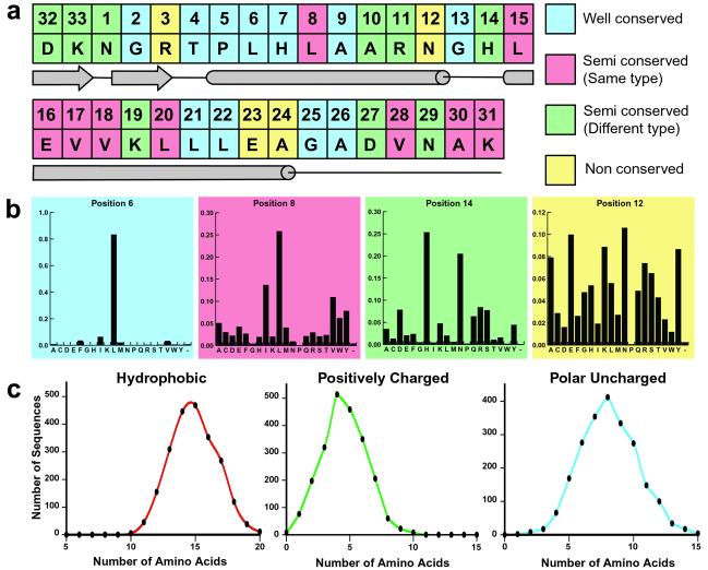
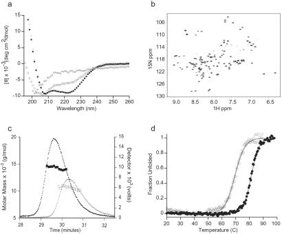
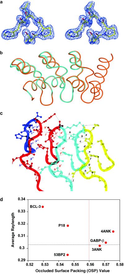
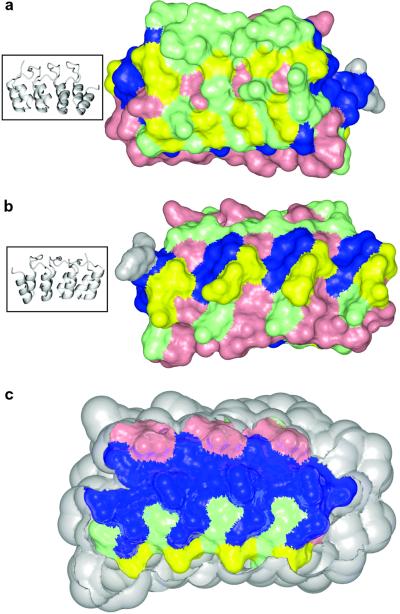
The ankyrin repeat is one of the most common, modular, protein-protein interaction motifs in nature. To understand the structural determinants of this family of proteins and extract the consensus information that defines the architecture of this motif, we have designed a series of idealized ankyrin repeat proteins containing one, two, three, or four repeats by using statistical analysis of approximately 4,000 ankyrin repeat sequences from the PFAM database. Biophysical and x-ray crystallographic studies of the three and four repeat constructs (3ANK and 4ANK) to 1.26 and 1.5 A resolution, respectively, demonstrate that these proteins are well-folded, monomeric, display high thermostability, and adopt a very regular, tightly packed ankyrin repeat fold. Mapping the degree of amino acid conservation at each position on the 4ANK structure shows that most nonconserved residues are clustered on the surface of the molecule that has been designated as the binding site in naturally occurring ankyrin repeat proteins. Thus, the consensus amino acid sequence contains all information required to define the ankyrin repeat fold. Our results suggest that statistical analysis and the consensus sequence approach can be used as an effective method to design proteins with complex topologies. These generic ankyrin repeat proteins can serve as prototypes for dissecting the rules of molecular recognition mediated by ankyrin repeats and for engineering proteins with novel biological functions.


Figures

Fig 1.
Fig 1.
Statistical analysis data used in the design of the consensus ankyrin repeat proteins. (a) Final sequence of the designed repeat and the corresponding secondary structure elements. Positions are colored according to the conservation level. (b) Representative histograms colored accordingly. (c) Examples of amino acid distribution data.
Fig 2.
Fig 2.
Solution characterization of the consensus ankyrin repeat proteins. (a) Far-UV CD spectra of 1ANK (⋄), 2ANK (□), (•), and (○) at 20°C. The pH values are described in the text. (b) 1H–15N HSQC spectra of recorded at 30°C, pH 4. (c) SEC–MALLS analysis of (○ and □) and (• and ▪). The detector values are the average of the 90° and 138° light scattering signals. The molar masses were calculated by using Astra software (Wyatt Technology, Santa Barbara, CA). (d) Thermal denaturation of (○) and (•) monitored by CD signals at 222 nm. The data were fit with an apparent two-state folding model (line).
Fig 3.
Fig 3.
X-ray crystal structures of and . (a) Stereo representation showing the initial electron density map of residues 37–40 of , including the TPLH motif, contoured at 1.5 σ. The figure was made with O and rendered by using MOLRAY (43). (b) Overlay of (cyan) and (orange) backbone structures. This figure and all following structural representations were made with MOLMOL (44). (c) Hydrogen bonding network in β-hairpin/loop region of . Each repeat is colored differently and hydrogen bonds are represented by letters: a, D32(OD1) and N34(HN); b, D32(O) and G35(HN); c, D32(OD1) and R36(HN); d, D32(HN) and R36(O); e, R36(O) and H7(HNE2); f, A30(O) and H7(HNE2); g, A30(HN) and D27(O); h, D27(OD1) and N29(HN); i, N29(OD1) and D60(HN); j, N29(ND2) and G58(O); k, N34(O) and K66(HN); and l, R36(HNE) and D65(OD2). Equivalent hydrogen bonds in adjacent repeats are represented by the letter followed by ′ or " and for all residue numbers i > 33, the canonical ankyrin repeat position = i − 33. (d) Comparison of intramolecular packing of ankyrin repeat proteins based on the occluded surface area analysis. For each protein, the average ray length of all residues was plotted against the average occluded surface packing (OSP) value of all buried residues with exposed surface area of <5%. The average values of 152 high-resolution structures in PDB are shown by the dashed lines on the graph (31).
Fig 4.
Fig 4.
Conservation level of residues mapped onto the structure of . Colors represent the conservation level: blue, well-conserved; pink, semiconserved of the same type; green, semiconserved of different type; and yellow, nonconserved. The C-terminal tyrosine is colored in gray. (a) Front (concave) surface of . (b) Back surface of . The ribbon diagrams on the left show orientations. (c) Longitudinal cross-section of the protein displaying the interior residues. Protein is in the same orientation as in b.

References

    1. Takahashi N., Takahashi, Y. & Putnam, F. W. (1985) Proc. Natl. Acad. Sci. USA 82, 1906-1910. - PMC - PubMed
    1. Sikorski R. S., Boguski, M. S., Goebl, M. & Hieter, P. (1990) Cell 60, 307-317. - PubMed
    1. Hirano T., Kinoshita, N., Morikawa, K. & Yanagida, M. (1990) Cell 60, 319-328. - PubMed
    1. Riggleman B., Weieschaus, E. & Schedl, P. (1989) Genes Dev. 3, 96-113. - PubMed
    1. Andrade M. A. & Bork, P. (1995) Nat. Genet. 11, 115-116. - PubMed

Publication types

LinkOut - more resources