. 2002 Dec 1;16(23):3074-86.
doi: 10.1101/gad.242602.

CD44 is required for two consecutive steps in HGF/c-Met signaling

Affiliations

CD44 is required for two consecutive steps in HGF/c-Met signaling

Véronique Orian-Rousseau et al. Genes Dev. .

Abstract

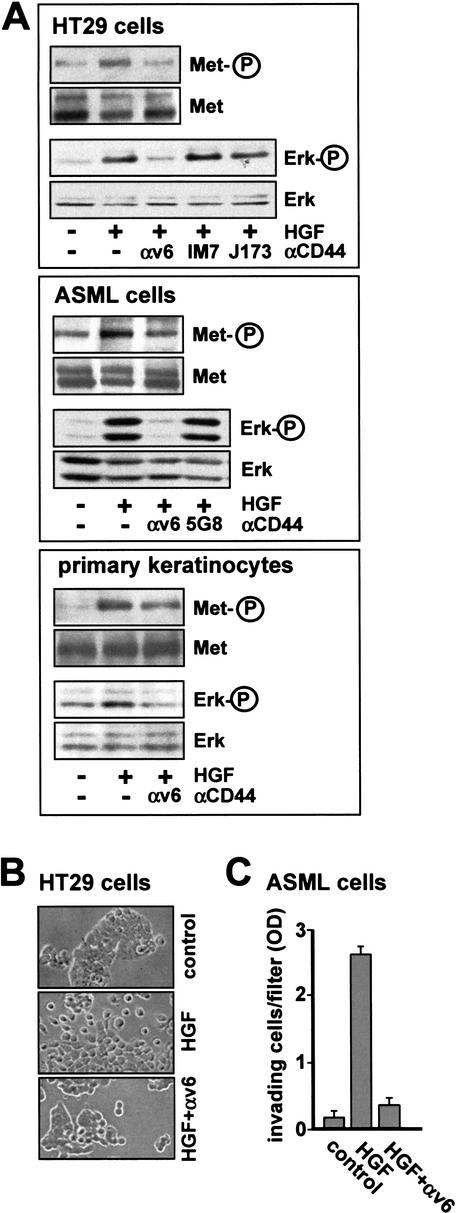
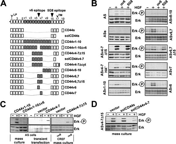
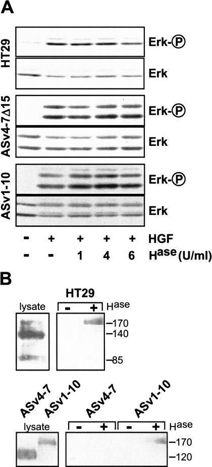
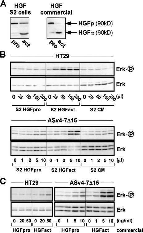
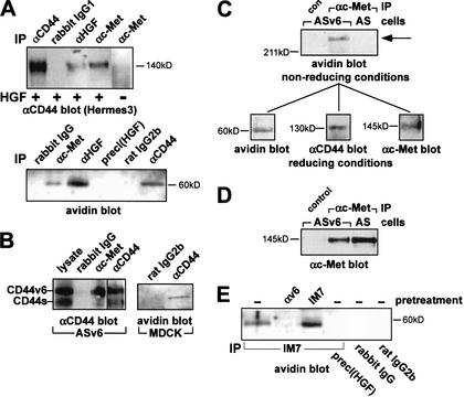
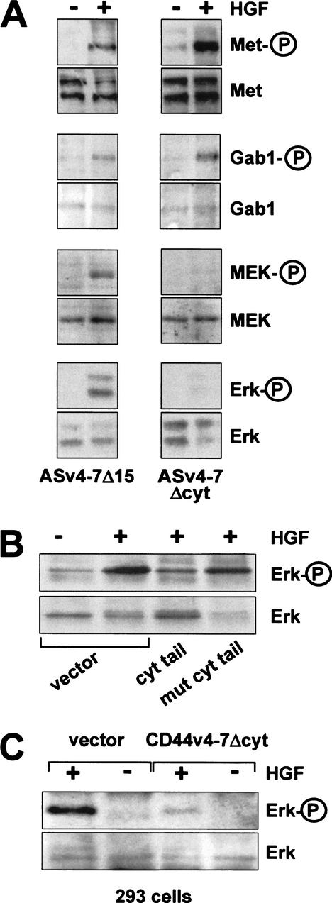
The tyrosine kinase receptor c-Met and its ligand HGF/SF, ezrin, and splice variants of CD44 have independently been identified as tumor metastasis-associated proteins. We now show that these proteins cooperate. A CD44 isoform containing variant exon v6 sequences is strictly required for c-Met activation by HGF/SF in rat and human carcinoma cells, in established cell lines as well as in primary keratinocytes. CD44v6-deficient tumor cells were unable to activate c-Met unless they were transfected with a CD44v6-bearing isoform. Antibodies to two v6-encoded epitopes inhibited autophosphorylation of c-Met by interfering with the formation of a complex formed by c-Met, CD44v6, and HGF/SF. In addition, signal transduction from activated c-Met to MEK and Erk required the presence of the cytoplasmic tail of CD44 including a binding motif for ERM proteins. This suggests a role for ERM proteins and possibly their link to the cortical actin cytoskeleton in signal transfer.


Figures

Figure 1
Figure 1
A CD44 variant isoform is required for Met activation. (A) HT29, ASML cells, and primary keratinocytes were each induced with HGF for 5 min in the presence or absence of antibodies (for HT29 cells and keratinocytes, αv6 was BIWA; for ASML cells αv6 was 1.1ASML). Subsequently, Met or Erk phosphorylations were determined (see Materials and Methods). (B) HT29 cells grown in the absence (control) or presence of HGF with or without antibodies directed against CD44v6 (BIWA) were visualized by phase contrast microscopy. Magnification, 40×. (C) Invasion of ASML cells into Matrigel was measured in the absence (control) or presence of HGF with or without antibodies directed against CD44v6 (1.1ASML). Each point represents the average of three independent determinations ±S.D.
Figure 2
Figure 2
Exon-specific RT–PCR analysis of CD44 isoforms in various cell lines. RT–PCR analyses with mRNA derived from the cells indicated were performed with CD44 exon-specific primers as described (van Weering et al. 1993; König et al. 1996). The marker lane (M) shows a 100-bp DNA ladder (Life Technologies).
Figure 3
Figure 3
CD44 isoforms containing the exon v6 sequence are required for Met activation. (A) Schematic representation of CD44 isoform structures. Constant region exons are numbered 1–5 and 15–19, variant exons v1–v10. LP, leader peptide; TM, transmembrane region. (B) Activation of Met in AS cells stably transfected with various CD44 isoforms was measured using phosphorylation of Erk as readout. The loading controls were performed by stripping the phospho-Erk blot and reprobing with an anti-Erk antibody. ASs indicates transfectants with CD44s, ASv4–7Δ15 cells stands for transfectants with CD44v4–7 with an exon 15 deletion, and ASv4–7HA indicates cells transfected with a CD44 isoform containing a point mutation in the major hyaluronate (HA) binding site (Sleeman et al. 1997). Other transfectants are designated according to the CD44 variants used for transfection. (C) CREF or AS cells were stably transfected with CD44 constructs (Günthert et al. 1991 and as indicated) together with a plasmid harboring the puromycin resistance gene, and were grown as mass cultures. AS cells were transiently transfected with CD44v4–v7 (or vector) together with a hemagglutinin-tagged Erk (see Materials and Methods). Determination of Erk activation in response to HGF treatment is described in Materials and Methods. (D) ASv4–7Δ15 cells were transfected with the indicated constructs, and puromycin resistant clones were selected as mass cultures.
Figure 4
Figure 4
Heparan sulfation of CD44 is not required for Met signaling. (A) HGF-induced Erk phosphorylation was measured in HT29, ASv4–7Δ15, and ASv1–10 cells. Where indicated, cells were pretreated with heparinase II (Hase) at 37°C for 3 h prior to HGF addition. (B) Western blot analyses using a ΔHS antibody are shown in the right panels. Where indicated, cells had been pretreated with heparinase II (6U/mL) at 37°C for 3 h. The lysates were probed by Western blotting using Hermes 3 (for HT29 cells) or 5G8 (for AS transfectants) antibodies.
Figure 5
Figure 5
HGF proform is activated by neither HT29 cells nor transfected AS cells. (A) HGF proform produced by Drosophila Schneider cells (S2 cells) as well as commercial pro-HGF were activated by incubation with 5% serum at 37°C overnight. HGF pro- and activated forms are visualized by Western blotting with anti-HGFα antibody. (B) Increasing amounts of conditioned medium from mock-transfected S2 cells (CM) or S2 cells transfected with an HGF expression vector and grown in the absence of serum (S2 HGFpro) or in the presence of 5% FCS (S2 HGFact) were used to treat HT29 cells or ASv4–7Δ15 cells. (C) Commercially available pro-HGF prior to and after activation (see A) was used to stimulate HT29 and ASv4–7Δ15 cells. We explain the slight induction of Erk phosphorylation at higher concentrations of HGF proform in the case of the very sensitive CD44v4–7-transfected AS cells by the presence of traces of activated HGF in the proform preparations.
Figure 6
Figure 6
CD44, Met, and HGF form a multimeric complex. (A) Western blots with anti-CD44 antibodies or with avidin of immunoprecipitates (IP) from HT29 cells treated with HGF where indicated, and cross-linked with DTSSP (see Materials and Methods; multimeric complex precipitations were also obtained without prior cross-linking; data not shown). For preclearing [precl(HGF)], lysates were immunoprecipitated with anti-HGF antibodies, and the supernatant was again immunoprecipitated with anti-HGFα. Rabbit IgG is a control antibody. (B) Western blots (5G8 antibodies) of immunoprecipitates from ASv6 cells (7.5% gel) and immunoprecipitates from MDCK cells using avidin for detection of biotinylated HGF (10% gel). (C) Immunoprecipitates of ASv6 or AS lysates using Met-specific antibodies were resolved under nonreducing conditions by SDS-PAGE (10%). A single band was identified that stained with either avidin (shown here) or antibodies directed against Met or CD44 (data not shown). The band was excised from the gel, incubated with dithiothreitol and rerun on SDS-PAGE. Western blots with avidin, 5G8 (αCD44), and α Met are shown. (D) Western blot for Met expression in lysates of ASv6 or AS cells. (E) Same procedure as in A. For immunoprecipitations, IM7 or control antibodies were used. Where indicated, cells were incubated with BIWA antibodies or IM7 at 37°C for 5 min prior to induction with biotinylated HGF.
Figure 7
Figure 7
The CD44 cytoplasmic tail is required for Met signaling, but not for Met activation. (A) In ASv4–7 cells or cells transfected with a cytoplasmic tail deletion (ASv4–7Δcyt), Met, Gab1, MEK, and Erk activation were determined. (B) Here, 293 cells were transiently cotransfected at a ratio of 1:15 with a hemagglutinin-tagged Erk construct and plasmids expressing GST fused to the cytoplasmic tail of CD44 or a mutant thereof, respectively (Morrison et al. 2001). Twenty-four hours after transfection, cells were starved for 10 h and then induced with HGF as indicated. Tagged Erk was immunoprecipitated, and phosphorylation of Erk was determined. (C) The ASv4–7Δcyt construct (or vector alone) was transiently transfected into 293 cells, and Erk activation upon HGF treatment was determined.

References

    1. Bardelli A, Comoglio PM. Scatter factor receptors are key players in a unique multistep program leading to invasive growth. Ciba Found Symp. 1997;212:133–144. - PubMed
    1. Bardelli A, Pugliese L, Comoglio PM. “Invasive-growth” signaling by the Met/HGF receptor: The hereditary renal carcinoma connection. Biochim Biophys Acta. 1997;1333:41–51. - PubMed
    1. Behrens J, Mareel MM, Van Roy FM, Birchmeier W. Dissecting tumor cell invasion: Epithelial cells acquire invasive properties after the loss of uvomorulin-mediated cell-cell adhesion. J Cell Biol. 1989;108:2435–2447. - PMC - PubMed
    1. Bennett KL, Jackson DG, Simon JC, Tanczos E, Peach R, Modrell B, Stamenkovic I, Plowman G, Aruffo A. CD44 isoforms containing exon v3 are responsible for the presentation of heparin-binding growth factor. J Cell Biol. 1995;128:687–698. - PMC - PubMed
    1. Birchmeier C, Gherardi E. Developmental roles of HGF/SF and its receptor, the c-Met tyrosine kinase. Trends Cell Biol. 1998;8:404–410. - PubMed

Publication types

LinkOut - more resources