

Review
doi: 10.1172/JCI16786.
Rescuing protein conformation: prospects for pharmacological therapy in cystic fibrosis
Affiliations
- PMID: 12464661
- PMCID: PMC151638
- DOI: 10.1172/JCI16786
Item in Clipboard
Review
Rescuing protein conformation: prospects for pharmacological therapy in cystic fibrosis
J Clin Invest.
2002 Dec.
No abstract available
Figures
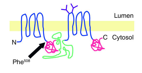
Cartoon representation of CFTR structure. Indicated are the transmembrane domains (blue), the two Asn-linked glycans (purple), the “R” domain (green), and the two nucleotide-binding domains (red). N, amino terminus; C, carboxy terminus.

Biogenesis and intracellular trafficking pathway of wild-type (a) and ΔF508 (b) CFTR. The width of the gray arrows is proportional to the relative flux through a particular branch of the pathway. Synthesis and cotranslational integration (step 1) in the ER membrane are followed by folding to a native conformation (step 2). About 25% of wild-type and more than 99% of ΔF508 CFTR molecules are degraded by a process that is mediated by cytoplasmic proteasomes (step 3). Native CFTR molecules (light blue cylinder) are delivered via the Golgi apparatus (not shown) to the plasma membrane (step 4), where they are subject to rapid endocytosis (step 5) to subapical vesicles (light blue lumen). CFTR is recycled to the plasma membrane (step 6), where it can be activated by cAMP-dependent kinases (step 7). Differences in the relative rates of recycling and degradation in lysosomes (pink lumen; step 8) are likely to account for the substantial differences in half-lives between wild-type and ΔF508 CFTR.

Following initial synthesis and membrane integration (step 1), nascent CFTR molecules are inefficiently folded (step 2) and are recognized as misfolded by one or more components of the quality control machinery (red; step 3a), which may interact with cytoplasmic, membrane, or lumenal portions of CFTR. This interaction is necessary to dislocate CFTR to the cytoplasmic ubiquitin (green spheres) conjugation machinery (step 3b), which then directs it to the proteasome for degradation (step 3c). Cytoplasmic aggregates form (step 3d) when the rate of production and dislocation of misfolded CFTR exceeds the capacity of the ubiquitin-proteasome system for degradation.
References
-
- Kerem B-S, et al. Identification of the cystic fibrosis gene: genetic analysis. Science. 1989;245:1073–1080. - PubMed
-
- Welsh MJ, Smith AE. Molecular mechanisms of CFTR chloride channel dysfunction in cystic fibrosis. Cell. 1993;73:1251–1254. - PubMed
-
- Kopito RR. Biosynthesis and degradation of CFTR. Physiol Rev. 1999;79(Suppl.):S167–S173. - PubMed
-
- Kleizen B, Braakman I, de Jonge HR. Regulated trafficking of the CFTR chloride channel. Eur J Cell Biol. 2000;79:544–556. - PubMed
Publication types
MeSH terms
Substances
LinkOut - more resources
Full Text Sources
Other Literature Sources
Medical
