

Increased production of IL-7 uncouples bone formation from bone resorption during estrogen deficiency
- PMID: 12464669
- PMCID: PMC151629
- DOI: 10.1172/JCI15687
Increased production of IL-7 uncouples bone formation from bone resorption during estrogen deficiency
Abstract
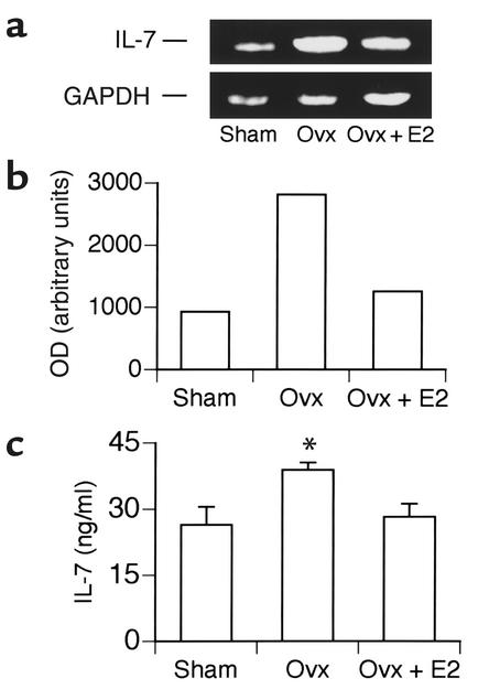
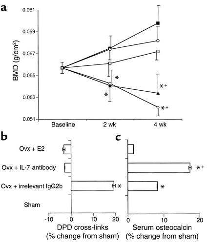
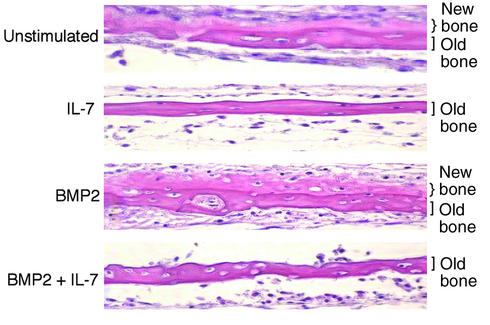
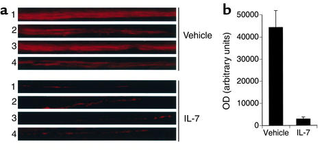
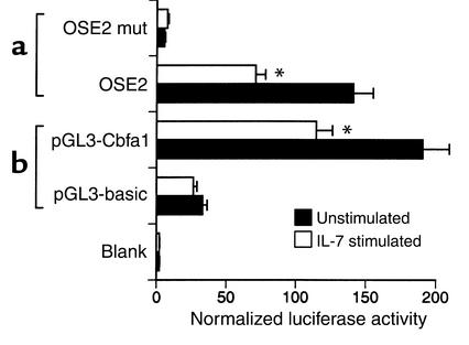
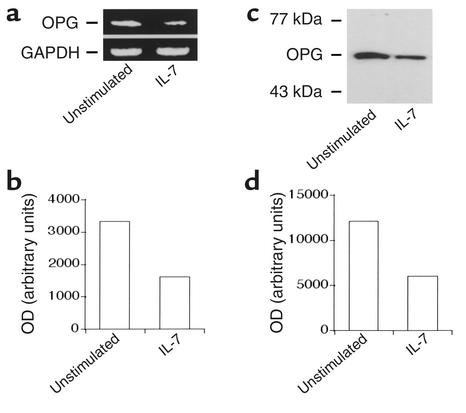
Postmenopausal bone loss stems from the inability of osteoblastic activity to match the increase in osteoclastic bone resorption induced by estrogen deficiency. However, the mechanism that uncouples osteoblast from osteoclast activities remains unexplained. We show that ovariectomy enhances the production of the osteoclastogenic cytokine IL-7, and that its neutralization in vivo prevents ovariectomy-induced bone loss. Surprisingly, serum osteocalcin levels, a biochemical marker of bone formation, suggested that the bone-sparing effects of IL-7 neutralization were due not only to inhibition of bone resorption, but also to stimulation of bone formation. Consistent with these data, addition of IL-7 to neonatal calvarial organ cultures blocked new bone formation, and injection of IL-7 into mice in vivo inhibited bone formation as measured by calcein incorporation into long bones. The antianabolic effects of IL-7 were consistent with an observed downregulation of the osteoblast-specific transcription factor core-binding factor alpha1/Runx2. Thus, because it targets both the osteoclast and the osteoblast pathways, IL-7 is central to the altered bone turnover characteristic of estrogen deficiency.
Figures
Similar articles
-
In vitro and ex vivo evidence that estrogens suppress increased bone resorption induced by ovariectomy or PTH stimulation through an effect on osteoclastogenesis.J Bone Miner Res. 1995 Oct;10(10):1523-30. doi: 10.1002/jbmr.5650101013. J Bone Miner Res. 1995. PMID: 8686508
-
Ablation of p38α MAPK Signaling in Osteoblast Lineage Cells Protects Mice From Bone Loss Induced by Estrogen Deficiency.Endocrinology. 2015 Dec;156(12):4377-87. doi: 10.1210/en.2015-1669. Epub 2015 Oct 6. Endocrinology. 2015. PMID: 26441240
-
Interleukin-1 receptor antagonist decreases bone loss and bone resorption in ovariectomized rats.J Clin Invest. 1994 May;93(5):1959-67. doi: 10.1172/JCI117187. J Clin Invest. 1994. PMID: 8182127 Free PMC article.
-
Estrogen regulation of immune cell bone interactions.Ann N Y Acad Sci. 2006 Apr;1068:256-74. doi: 10.1196/annals.1346.030. Ann N Y Acad Sci. 2006. PMID: 16831927 Review.
-
Estrogen, cytokines, and the control of osteoclast formation and bone resorption in vitro and in vivo.Osteoporos Int. 1993;3 Suppl 1:114-6. doi: 10.1007/BF01621882. Osteoporos Int. 1993. PMID: 8461536 Review. No abstract available.
Cited by
-
Advances in the research on myokine-driven regulation of bone metabolism.Heliyon. 2023 Nov 20;10(1):e22547. doi: 10.1016/j.heliyon.2023.e22547. eCollection 2024 Jan 15. Heliyon. 2023. PMID: 38226270 Free PMC article. Review.
-
Estrogen deficiency and bone loss: an inflammatory tale.J Clin Invest. 2006 May;116(5):1186-94. doi: 10.1172/JCI28550. J Clin Invest. 2006. PMID: 16670759 Free PMC article. Review.
-
Microbial osteoporosis: The interplay between the gut microbiota and bones via host metabolism and immunity.Microbiologyopen. 2019 Aug;8(8):e00810. doi: 10.1002/mbo3.810. Epub 2019 Apr 18. Microbiologyopen. 2019. PMID: 31001921 Free PMC article. Review.
-
Multiple Myeloma and Bone: The Fatal Interaction.Cold Spring Harb Perspect Med. 2018 Aug 1;8(8):a031286. doi: 10.1101/cshperspect.a031286. Cold Spring Harb Perspect Med. 2018. PMID: 29229668 Free PMC article. Review.
-
Immunology of Gut-Bone Signaling.Adv Exp Med Biol. 2017;1033:59-94. doi: 10.1007/978-3-319-66653-2_5. Adv Exp Med Biol. 2017. PMID: 29101652 Free PMC article.
References
-
- Manolagas SC, Jilka RL. Bone marrow, cytokines, and bone remodeling. N Engl J Med. 1995;332:305–311. - PubMed
-
- Hofmeister R, et al. Interleukin-7: physiological roles and mechanisms of action. Cytokine Growth Factor Rev. 1999;10:41–60. - PubMed
-
- Fry TJ, Mackall CL. Interleukin-7: from bench to clinic. Blood. 2002;99:3892–3904. - PubMed
-
- Weitzmann MN, Cenci S, Rifas L, Brown C, Pacifici R. Interleukin-7 stimulates osteoclast formation by up-regulating the T-cell production of soluble osteoclastogenic cytokines. Blood. 2000;96:1873–1878. - PubMed
Publication types
MeSH terms
Substances
Grants and funding
LinkOut - more resources
Full Text Sources
Other Literature Sources
