. 2002 Dec;110(11):1667-74.
doi: 10.1172/JCI16991.

A monoclonal thyroid-stimulating antibody

Affiliations

A monoclonal thyroid-stimulating antibody

Takao Ando et al. J Clin Invest. 2002 Dec.

Abstract

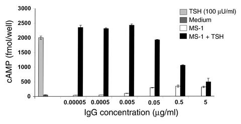
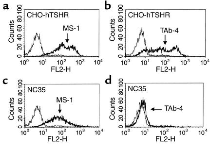
The thyrotropin receptor, also known as the thyroid-stimulating hormone receptor (TSHR), is the primary antigen of Graves disease. Stimulating TSHR antibodies are the cause of thyroid overstimulation and were originally called long-acting thyroid stimulators due to their prolonged action. Here we report the successful cloning and characterization of a monoclonal antibody (MS-1) with TSHR-stimulating activity. The thyroid-stimulating activity of MS-1 was evident at IgG concentrations as low as 20 ng/ml. MS-1 also competed for radiolabeled TSH binding to the native TSHR and was able to compete for TSH-induced stimulation. MS-1 recognized a conformational epitope within the TSHR alpha (or A) subunit but excluding the receptor cleavage region. Using an assay measuring loss of antibody recognition after cleavage we demonstrated that MS-1, in contrast to TSH, was unable to enhance TSHR posttranslational cleavage. Since receptor cleavage is followed by alpha subunit shedding and receptor degradation, the functional half-life of the receptor may be extended. The isolation and characterization of MS-1 provides a novel explanation for the prolonged thyroid stimulation in this disease which may be secondary to the lack of receptor cleavage in addition to the prolonged half-life of IgG itself.


Figures

Figure 1
Figure 1
cAMP dose-response study of TSHR-stimulating monoclonal antibody MS-1. MS-1 was examined for thyroid-stimulating activity at the indicated concentrations. TAb-4 and TAb-8 (5 μg/ml) were used as negative controls. Thyroid stimulation was expressed as a ratio with basal cAMP levels induced by culture medium. More than 200% stimulation above baseline was judged as positive and is shown as a dotted line.
Figure 2
Figure 2
Dose-response study of the blocking activity of MS-1. CHO-hTSHR cells were stimulated with MS-1 at the indicated concentrations with or without 100 μU/ml of TSH. Since MS-1 is a TSHR-stimulating monoclonal antibody, the cAMP generated by MS-1 alone was subtracted from the cAMP generated by MS-1 plus TSH. Medium alone produced less than 50 fmol/well cAMP, and TSH (100 μU/ml) generated 2,000 fmol/well.
Figure 3
Figure 3
Recognition of a TSHR conformational epitope by MS-1. MS-1 recognized the native conformation of hTSHR antigen on unfixed CHO-hTSHR cells (a), but not on fixed CHO-hTSHR cells (b). In contrast to MS-1, mouse monoclonal TSHR-Ab RSR-1 (29) recognized both the native (c) and fixed (d) conformations of the hTSHR. Thin and thick lines indicate staining on CHO and CHO-hTSHR cells, respectively. Fluorescence intensity was measured in FL-1.
Figure 4
Figure 4
Epitope recognition within TSHR residues 317–366 by TAb-4, but not MS-1. MS-1 reacted similarly to both CHO-hTSHR (a) and NC35 (c) cells. However, strong immunoreactivity seen on CHO-hTSHR cells (b) by TAb-4 was lost on NC35 cells (d) lacking TSHR residues 317–366. The thick lines indicate CHO-hTSHR cells in a and b, and NC35 cells in c and d. The thin lines indicate CHO cells. Fluorescence intensity was measured in FL-2.
Figure 5
Figure 5
TSHR cleavage assay. CHO-hTSHR cells were treated with increasing concentrations of TSH for 24 hours and stained with two monoclonal TSHR-Ab’s in order to examine the effect of TSH on TSHR cleavage. RSR-1 (white bars) recognized the β subunit of TSHR, in contrast to the recognition of the region subjected to receptor cleavage (residues 317–366) by RSR-4 (black bars). It should be noted that RSR-4 had no TSH-competing activity. Reduction of immunoreactivity by RSR-4 was a measure of TSHR cleavage and was observed after treatment with TSH (more than 10 μU/ml).
Figure 6
Figure 6
TSHR cleavage is not induced by MS-1. CHO-hTSHR cells were treated with 100 μU/ml of TSH for 24 hours and stained with two monoclonal TSHR-Ab’s, and showed a reduction in the immunoreactivity of RSR-4 (a) but not RSR-1 (b). In contrast, similar treatment with MS-1 (5 μg/ml) did not alter immunoreactivity by either RSR-4 (c) or RSR-1 (d), indicating that MS-1 did not accelerate TSHR cleavage. The thin lines indicate staining of CHO-hTSHR cells without TSH (a and b) or MS-1 (c and d), and thick lines indicate CHO-hTSHR cells with TSH (a and b) and MS-1 (c and d), respectively.

References

    1. Davies T, Marians R, Latif R. The TSH receptor reveals itself. J Clin Invest. 2002;110:161–164. doi:10.1172/JCI200216234. - PMC - PubMed
    1. Loosfelt H, et al. Two-subunit structure of the human thyrotropin receptor. Proc Natl Acad Sci USA. 1992;89:3765–3769. - PMC - PubMed
    1. Rees Smith B, McLachlan SM, Furmaniak J. Autoantibodies to the thyrotropin receptor. Endocr Rev. 1988;9:106–121. - PubMed
    1. Chazenbalk GD, et al. Evidence that the thyrotropin receptor ectodomain contains not one, but two, cleavage sites. Endocrinology. 1997;138:2893–2899. - PubMed
    1. de Bernard S, et al. Sequential cleavage and excision of a segment of the thyrotropin receptor ectodomain. J Biol Chem. 1999;274:101–107. - PubMed

Publication types