. 2002 Dec;110(11):1703-16.
doi: 10.1172/JCI15849.

Critical role for CXCR2 and CXCR2 ligands during the pathogenesis of ventilator-induced lung injury

Affiliations

Critical role for CXCR2 and CXCR2 ligands during the pathogenesis of ventilator-induced lung injury

John A Belperio et al. J Clin Invest. 2002 Dec.

Abstract

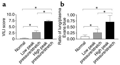
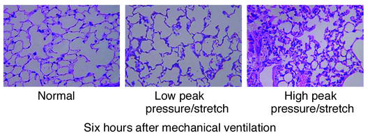
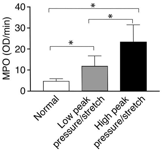
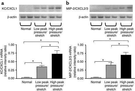
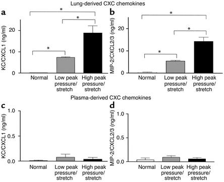
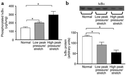
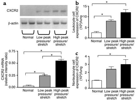
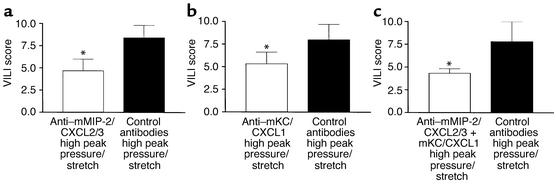
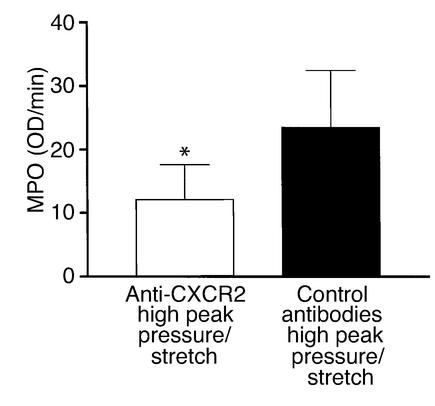
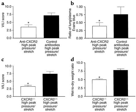
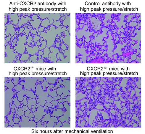
Mortality related to adult respiratory distress syndrome (ARDS) ranges from 35% to 65%. Lung-protective ventilator strategies can reduce mortality during ARDS. The protective strategies limit tidal volumes and peak pressures while maximizing positive end-expiratory pressure. The efficacy of this approach is due to a reduction of shear-stress of the lung and release of inflammatory mediators. Ventilator-induced lung injury (VILI) is characterized by inflammation. The specific mechanism(s) that recruit leukocytes during VILI have not been elucidated. Because the murine CXC chemokines KC/CXCL1 and MIP-2/CXCL2/3, via CXCR2, are potent neutrophil chemoattractants, we investigated their role in a murine model of VILI. We compared two ventilator strategies in C57BL/6 mice: high peak pressure and high stretch (high peak pressure/stretch) versus low peak pressure/stretch for 6 hours. Lung injury and neutrophil sequestration from the high-peak pressure/stretch group were greater than those from the low-peak pressure/stretch group. In addition, lung expression of KC/CXCL1 and MIP-2/CXCL2/3 paralleled lung injury and neutrophil sequestration. Moreover, in vivo inhibition of CXCR2/CXC chemokine ligand interactions led to a marked reduction in neutrophil sequestration and lung injury. These findings were confirmed using CXCR2(-/-) mice. Together these experiments support the notion that increased expression of KC/CXCL1 and MIP-2/CXCL2/3 and their interaction with CXCR2 are important in the pathogeneses of VILI.


Figures

Figure 1
Figure 1
VILI. (a) VILI scores by analysis of H&E-stained histopathologic sections from mouse lungs after 6 hours of mechanical ventilation. A cumulative score was based on leukocyte infiltration, exudative edema, hemorrhage, and alveolar wall thickness (n = 15; three random sections per lung and five lungs per group). *P < 0.02. (b) Evans blue permeability index where Evans blue was given 3 hours after the start of mechanical ventilation and mice were sacrificed at the 6-hour time point (n = 10 lungs per group). *P < 0.02.
Figure 2
Figure 2
Histopathology of lungs is associated with mechanical ventilation. Representative photomicrographs with H&E staining (×400) of the lung histopathology from mice placed on either high– or low–peak pressure/stretch mechanical ventilation, or from normal nonventilated mice.
Figure 3
Figure 3
Lung neutrophil sequestration as demonstrated by MPO levels from mice placed on either high– or low–peak pressure/stretch mechanical ventilation, or from normal nonventilated mice (n = 10 mice per group). *P < 0.02.
Figure 4
Figure 4
KC/CXCL1 and MIP-2/CXCL2/3 mRNA levels are markedly elevated during VILI. (a) RT-PCR determination of KC/CXCL1 mRNA in lung homogenates from mice placed on either high– or low–peak pressure/stretch ventilator protocol, or from normal nonventilated mice. Semiquantitative results are expressed as a ratio of each PCR product to β-actin band density (n = 4 mice per group). *P < 0.02. (b) RT-PCR determination of MIP-2/CXCL2/3 mRNA in lung homogenates from mice placed on either high– or low–peak pressure/stretch ventilator protocol, or from normal nonventilated mice. Semiquantitative results are expressed as a ratio of each PCR product to β-actin band density (n = 4 mice per group). *P < 0.02.
Figure 5
Figure 5
KC/CXCL1 and MIP-2/CXCL2/3 protein levels are markedly elevated during VILI. (a and b) ELISA measurements of (a) KC/CXCL1 protein levels and (b) MIP-2/CXCL2/3 protein levels in lung homogenates from mice placed on either high– or low–peak pressure/stretch ventilator protocol, or from normal nonventilated mice (n = 10 mice per group). *P < 0.02. (c and d) ELISA measurements of (c) KC/CXCL1 protein levels and (d) MIP-2/CXCL2/3 protein levels in plasma from mice placed on either high– or low–peak pressure/stretch ventilator protocol, or from normal nonventilated mice (n = 10 mice per group). P < 0.02.
Figure 6
Figure 6
There is increased phosphorylation of IκBα and more degradation of IκBα during the pathogenesis of VILI. (a) Measurements of phosphorylation state of IκBα in whole-lung homogenates from mice placed on either high– or low–peak pressure/stretch ventilator protocol for 2 hours, or from normal nonventilated mice (n = 6 mice per group). *P < 0.02. (b) Western blot analysis of IκBα protein levels in whole-lung homogenates from mice placed on either high– or low–peak pressure/stretch ventilator protocol for 2 hours, or from normal nonventilated mice. Semiquantitative results are expressed as a band density area (n = 6 mice per group). *P < 0.02.
Figure 7
Figure 7
(a) CXCR2 mRNA expression is markedly elevated during VILI. RT-PCR determination of CXCR2 mRNA in lung homogenates from mice placed on either high– or low–peak pressure/stretch ventilator protocol, or from normal nonventilated mice. Semiquantitative results are expressed as a ratio of each PCR product to β-actin band density (n = 4 mice per group). *P < 0.02. (b and c) FACS analysis of CXCR2. (b) Total number of leukocytes (CD45-positive cells) with cell surface expression of CXCR2 in whole-lung digest homogenates from mice placed on either high– or low–peak pressure/stretch ventilator protocol, or from normal nonventilated mice (n = 3 mice per group). *P < 0.02. (c) Total number of nonleukocytes (CD45-negative cells) with cell surface expression of CXCR2 in whole-lung digest homogenates from mice placed on either high– or low–peak pressure/stretch ventilator protocol, or from normal nonventilated mice (n = 3 mice per group). *P < 0.02.
Figure 8
Figure 8
In vivo neutralization of (a) mMIP-2/CXCL2/3, (b) mKC/CXCL1, and (c) mKC/CXCL1 + mMIP-2/CXCL2/3 (as compared with treatment with control antibodies) attenuates VILI, as demonstrated by VILI scores of H&E-stained histopathologic sections from mice placed on high peak pressure/stretch. A cumulative score was based on leukocyte infiltration, exudative edema, hemorrhage, and alveolar wall thickness (n = 6; two random sections per lung and three lungs per group). *P < 0.05.
Figure 9
Figure 9
In vivo neutralization of murine CXCR2 (as compared with treatment with control antibodies) attenuates lung neutrophil sequestration as determined by MPO levels from mice placed on a high–peak pressure/stretch ventilator protocol (n = 10 mice per group). *P < 0.05.
Figure 10
Figure 10
In vivo neutralization of murine CXCR2 attenuates VILI. (a) VILI scores of H&E-stained histopathologic sections from mice placed on high–peak pressure/stretch ventilation protocol treated with neutralizing antibodies to CXCR2 or with control antibodies. A cumulative score was based on leukocyte infiltration, exudative edema, hemorrhage, and alveolar wall thickness (n = 15; three random sections per lung and five lungs per group). *P < 0.05. (b) Evans blue permeability index (n = 10 mice per group). *P < 0.05. (c) VILI scores of H&E-stained histopathologic sections from CXCR2–/– mice placed on high–peak pressure/stretch ventilation protocol, as compared with CXCR2+/+ mice (n = 15; three random sections per lung and five lungs per group). *P < 0.05. (d) Wet-to-dry weight ratio of lungs from CXCR2–/– and CXCR2+/+ mice placed on high–peak pressure/stretch ventilation protocol (n = 8 mice per group). *P < 0.05.
Figure 11
Figure 11
Histopathology of lungs is associated with mechanical ventilation. Representative photomicrographs (×400) with H&E staining of the lung histopathology. (a) Mice placed on high–peak pressure/stretch mechanical ventilation treated with anti–murine CXCR2 or with control antibodies. (b) CXCR2–/– mice placed on high–peak pressure/stretch mechanical ventilation, as compared with CXCR2+/+ mice.

Comment in

References

    1. Ware LB, Matthay MA. The acute respiratory distress syndrome. N Engl J Med. 2000;342:1334–1349. - PubMed
    1. Ranieri VM, et al. Effect of mechanical ventilation on inflammatory mediators in patients with acute respiratory distress syndrome: a randomized controlled trial. JAMA. 1999;282:54–61. - PubMed
    1. Kollef MH, Schuster DP. The acute respiratory distress syndrome. N Engl J Med. 1995;332:27–37. - PubMed
    1. Dreyfuss D, Soler P, Basset G, Saumon G. High inflation pressure pulmonary edema. Respective effects of high airway pressure, high tidal volume, and positive end-expiratory pressure. Am Rev Respir Dis. 1988;137:1159–1164. - PubMed
    1. Corbridge TC, et al. Adverse effects of large tidal volume and low PEEP in canine acid aspiration. Am Rev Respir Dis. 1990;142:311–315. - PubMed

Publication types