. 2002 Dec;110(11):1739-47.
doi: 10.1172/JCI15695.

T cell activation causes diarrhea by increasing intestinal permeability and inhibiting epithelial Na+/K+-ATPase

Affiliations

T cell activation causes diarrhea by increasing intestinal permeability and inhibiting epithelial Na+/K+-ATPase

Mark W Musch et al. J Clin Invest. 2002 Dec.

Abstract

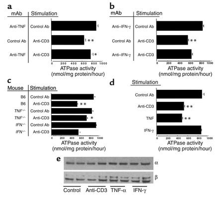
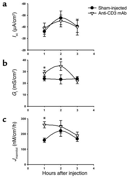
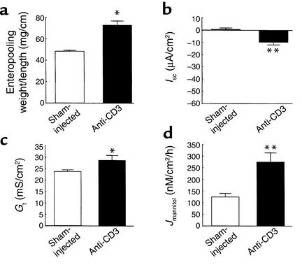
Inflammatory bowel disease (IBD) is associated with mucosal T cell activation and diarrhea. We found that T cell activation with anti-CD3 mAb induces profound diarrhea in mice. Diarrhea was quantified by intestinal weight-to-length (wt/l) ratios, mucosal Na(+)/K(+)-ATPase activity was determined and ion transport changes were measured in Ussing chambers. Anti-CD3 mAb increased jejunal wt/l ratios by more than 50% at 3 hours, returning to base line after 6 hours. Fluid accumulation was significantly reduced in TNF receptor-1 (TNFR-1(-/-)), but not IFN-gamma knockout mice. Anti-CD3 mAb decreased mucosal Na(+)/K(+)-ATPase activity, which was blocked by anti-TNF mAb and occurred to a lesser degree in TNFR-1(-/-) mice. Neither alpha nor beta subunits of Na(+)/K(+)-ATPase decreased in abundance at 3 hours. Intestinal tissue from anti-CD3-treated mice exhibited increased permeability to mannitol at 1 hour and decreases in electroneutral Na(+) absorption, Na(+)-dependent glucose absorption, and cAMP-stimulated anion secretion at 3 hours. Furthermore, enteral fluid accumulation was observed in CFTR(-/-) mice, indicating a minor role of active anion secretion. These data suggest that diarrhea in IBD is due to TNF-mediated malabsorption rather than to secretory processes. T cell activation induces luminal fluid accumulation by increasing mucosal permeability and reducing epithelial Na(+)/K(+)-ATPase activity leading to decreased intestinal Na(+) and water absorption.


Figures

Figure 1
Figure 1
(a) Time course of effect of anti-CD3 mAb on jejunal wt/l ratio. Mice were injected with 0.2 mg anti-CD3 mAb at time 0. Loop wt/l ratios were measured after varying times. Values are means ± SE for six determinations. (b) Effect of anti-TNF and anti–IFN-γ antibodies on jejunal wt/l ratios at 3 hours. Mice were injected simultaneously with 0.2 mg control or anti-CD3 mAb along with neutralizing mAb to TNF or IFN-γ. (c) Control or anti-CD3 mAb was injected in C57BL/6 or in TNFR-1 or IFN-γ knockout mice. Weight-to-length ratios were determined after 3 hours. Values are means ± SE for six determinations in each group. In all cases, *P < 0.05, **P < 0.01 by comparison using ANOVA. In (a), all comparisons were with 0-time control. In (b) and (c), control mAb-treated, anti–CD3-stimulated mice were initially compared with control mAb-treated, unstimulated controls. In (b), results in anti-TNF mAb and anti–IFN-γ–treated, anti–CD3-stimulated mice were compared with control mAb-treated, anti–CD3-stimulated mice. In (c), results in anti–CD3-stimulated TNFR-1–/– and IFN-γ–/– mice were compared with anti–CD3-stimulated B6 mice. The data indicate that TNF but not IFN-γ inhibition prevented anti–CD3-induced diarrhea.
Figure 2
Figure 2
Effect of TNF or IFN-γ on wt/l ratios. (a) Mice were injected with control antibody, anti-CD3 mAb, TNF, or IFN-γ. Jejunal wt/l ratios were measured at 3 hours. Values are means ± SE for six determinations. (b) Dose-dependence of TNF effect. Mice were injected with varying amounts of TNF, and jejunal wt/l ratios were measured at 3 hours. Values are means ± SE for six determinations in each group or each TNF dose. *P < 0.05, **P < 0.01 compared with control antibody in a and 0 TNF in b by ANOVA.
Figure 3
Figure 3
(ad) Effect of anti-CD3 mAb on Na+/K+-ATPase activity. Mice were injected with control or anti-CD3 mAb and enzyme activity measured in jejunum isolated 3 hours later. Values are means ± SE for four to six determinations in each group. Results in unstimulated mice treated with anti-TNF (a) or anti–IFN-γ (b) mAbs were not different from control mAb-treated, unstimulated mice (data not shown). In (a) and (b), results in control mAb-treated, anti–CD3-stimulated mice were compared with unstimulated mice. Results of anti–cytokine-treated, anti–CD3-stimulated mice were compared with control mAb-treated, anti–CD3-stimulated mice. In (c), data in B6, anti–CD3-stimulated mice were compared with B6, unstimulated mice and results in anti-CD3-stimulated, TNFR-1–/– and IFN-γ–/– mice were compared with B6, anti–CD3-stimulated mice. In (d), results in anti-CD3 mAb-stimulated, TNF-treated and IFN-γ–treated mice were compared with control mAb-treated mice. (e) Effect of anti-CD3 mAb on abundance of Na+/K+-ATPase α and β subunits. Microsomal membranes were isolated and analyzed on SDS-PAGE, and Western blots were performed using mAb to the α and β subunits. *P < 0.05, **P < 0.01 compared with control by ANOVA.
Figure 4
Figure 4
Time course of effect of anti-CD3 mAb on bioelectric parameters and intestinal permeability of murine jejunum. Mice were injected with PBS (Sham-injected) or 0.2 mg anti-CD3 mAb at time 0. Jejunal sections were mounted in Ussing chambers for measurement of Isc, Gt, and Jmannitol. Values are means ± SE for 15 sections (eight mice) at 1 hour, eight sections (five mice) at 2 hours, and eight sections (four mice) at 3 hours. *P < 0.05, **P < 0.1 vs. sham-injected.
Figure 5
Figure 5
Effect of anti-CD3 mAb on Na+ absorption and anion secretion across murine jejunum. Mice were injected with PBS (Sham-injected) or 0.2 mg anti-CD3 mAb 3 hours before sacrifice. Jejunal sections were mounted in Ussing chambers for measurement of Isc and 22Na flux measurements. (a) Mucosal-to-serosal 22Na flux sensitive to 100 μM EIPA (n = 10 sections from six mice). (b) Isc response to addition of 10 mM glucose in mucosal bath (15 minutes) to stimulate Na+-coupled glucose transport (n = 12 sections from six mice). (c) Isc response to addition of 10 μM forskolin in bathing solutions (10 minutes) to stimulate cAMP-dependent anion secretion (n = 11 sections from six mice). Values are means ± SE. *P < 0.05, **P < 0.1 vs. sham-injected.
Figure 6
Figure 6
Effect of anti-CD3 mAb in CFTR-deficient mice. Mice were injected with PBS (Sham-injected) or 0.2 mg anti-CD3 mAb 3 hours before sacrifice. (a) Effect on jejunal wt/l ratio. Values are means ± SE for three mice in each group. *P < 0.5 vs. sham-injected. (bd) Effect of anti-CD3 mAb ISC, Gt and Jmannitol of intestine from CFTR-deficient murine jejunum. Values are means ± SE for seven sections (four mice). *P < 0.05, **P < 0.1 vs. sham-injected.

References

    1. Ciancio MJ, Chang EB. Epithelial secretory response to inflammation. Ann NY Acad Sci. 1992;664:210–221. - PubMed
    1. Baert FJ, et al. Tumor necrosis factor-alpha antibody (Infliximab) therapy profoundly down-regulates the inflammation in Crohn’s ileocolitis. Gastroenterology. 1999;116:22–28. - PubMed
    1. Bell SJ, Kamm MA. Antibodies to tumor necrosis factor alpha as treatment for Crohn’s disease. Lancet. 2000;355:858–860. - PubMed
    1. van Dullemen HM, et al. Treatment of Crohn’s disease with anti-tumor necrosis factor chimeric monoclonal antibody (cA2) Gastroenterology. 1995;109:129–135. - PubMed
    1. Casellas F, et al. Intraluminal colonic release of immunoreactive tumour necrosis factor in chronic ulcerative colitis. Clin Sci (Lond) 1994;87:453–458. - PubMed

Publication types

MeSH terms