. 2002 Dec;161(6):2153-67.
doi: 10.1016/S0002-9440(10)64493-8.

Nipah virus infection: pathology and pathogenesis of an emerging paramyxoviral zoonosis

Affiliations

Nipah virus infection: pathology and pathogenesis of an emerging paramyxoviral zoonosis

Kum Thong Wong et al. Am J Pathol. 2002 Dec.

Abstract

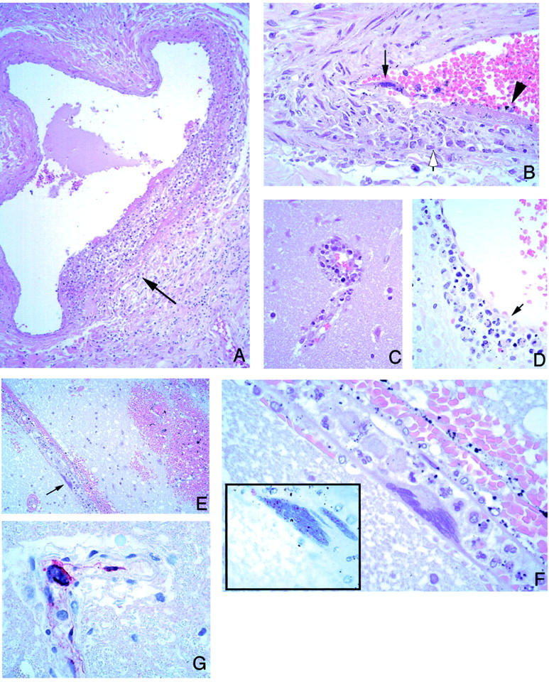
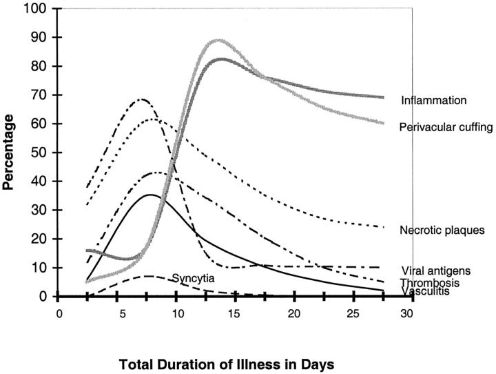
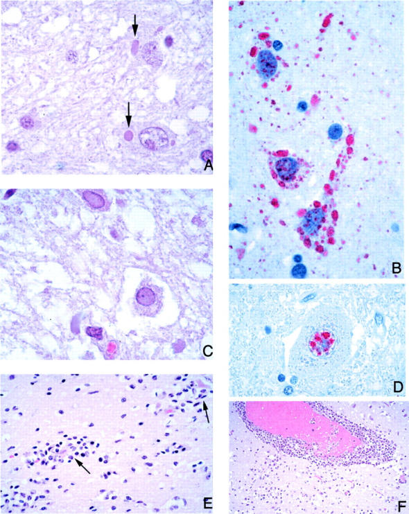
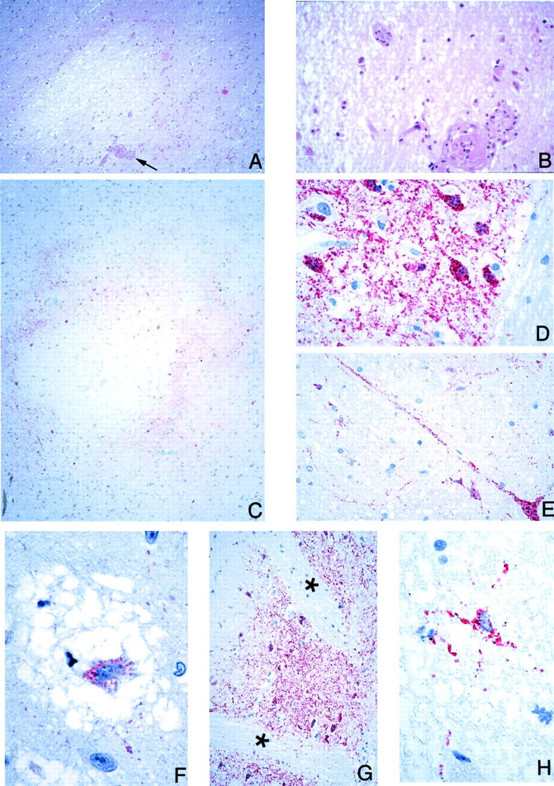
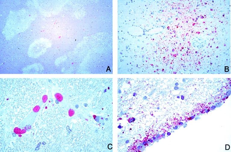
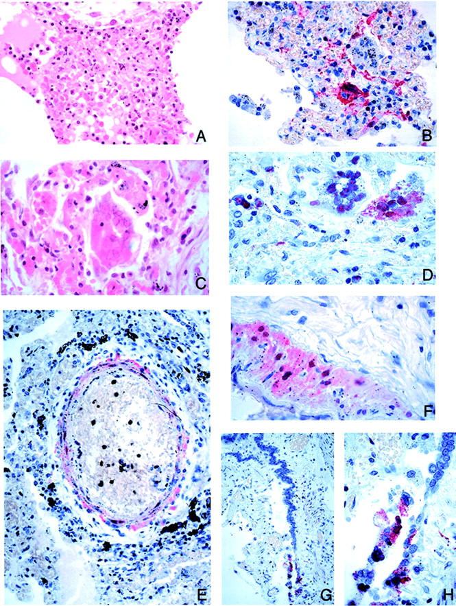
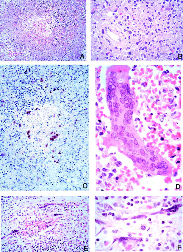
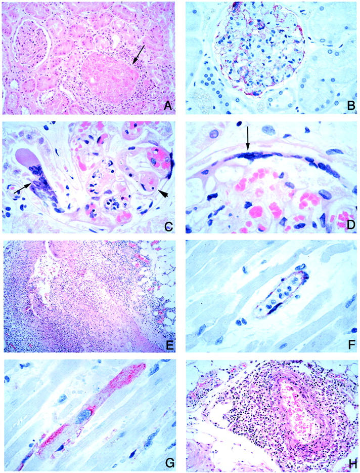
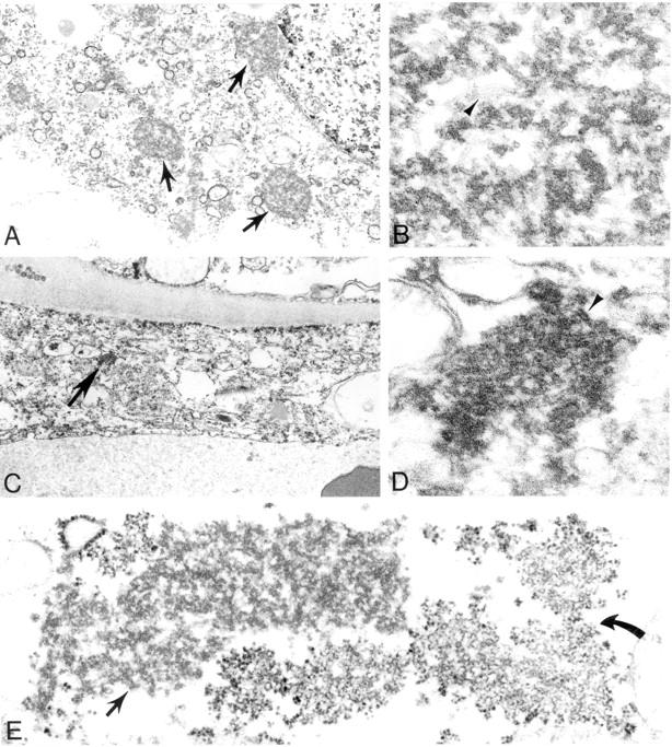
In 1998, an outbreak of acute encephalitis with high mortality rates among pig handlers in Malaysia led to the discovery of a novel paramyxovirus named Nipah virus. A multidisciplinary investigation that included epidemiology, microbiology, molecular biology, and pathology was pivotal in the discovery of this new human infection. Clinical and autopsy findings were derived from a series of 32 fatal human cases of Nipah virus infection. Diagnosis was established in all cases by a combination of immunohistochemistry (IHC) and serology. Routine histological stains, IHC, and electron microscopy were used to examine autopsy tissues. The main histopathological findings included a systemic vasculitis with extensive thrombosis and parenchymal necrosis, particularly in the central nervous system. Endothelial cell damage, necrosis, and syncytial giant cell formation were seen in affected vessels. Characteristic viral inclusions were seen by light and electron microscopy. IHC analysis showed widespread presence of Nipah virus antigens in endothelial and smooth muscle cells of blood vessels. Abundant viral antigens were also seen in various parenchymal cells, particularly in neurons. Infection of endothelial cells and neurons as well as vasculitis and thrombosis seem to be critical to the pathogenesis of this new human disease.


Figures

Figure 1.
Figure 1.
Vascular pathology and viral immunolocalization in Nipah virus infection. A: Vasculitis in a lung artery. Note focal and transmural mixed inflammatory infiltrate (arrow). B: High-power magnification of another pulmonary vessel showing endothelial syncytium (arrow) and ulceration (arrowhead). Note focal transmural inflammation (open arrow). C: Vasculitis in a small cerebral vessel. D: A cerebral venule showing endothelial ulceration (arrow) associated with inflammatory cellular debris. E: Cerebral hemorrhage adjacent to a vessel with a multinucleated giant cell (arrow). F: Higher-power magnification showing endothelial origin of syncytium seen in E. Inset: Intranuclear immunostaining of viral antigens in same cell. G: Positive immunostaining is also seen in the cytoplasm of an endothelial syncytial cell protruding in lumen of a cerebral vessel. H&E stain: A-F and inset in F; immunoalkaline phosphatase with napthol fast red substrate and hematoxylin counterstain, G. Original magnifications: ×25 (A); ×100 (B-D); ×50 (E); ×250 (F, inset); ×158 (F, G).
Figure 2.
Figure 2.
Temporal distribution of microscopic findings and viral antigen in the CNS in fatal Nipah virus infection. Cases were grouped into four subgroups by duration of illness: 0 to 5 days (n = 6), 6 to 10 days (n = 18), 11 to 15 days (n = 3), and 16 to 35 days (n = 4). The percentage of a particular histopathological lesion found in each subgroup was calculated by using as the numerator the number of CNS sections with at least one positive lesion found therein. The denominator is the total number of CNS sections in that subgroup. Because the duration of illness for case 32 (Table 1) ▶ could not be determined with accuracy, it was excluded.
Figure 3.
Figure 3.
CNS pathology and viral immunolocalization in Nipah virus infection. A: Typical eosinophilic viral inclusions in cytoplasm of several neurons (arrows). B: The viral nature of these inclusions is evidenced by immunostaining of Nipah viral antigens. C: Less-common intranuclear neuronal inclusions occupying most of the nucleus and pushing the chromatin to the periphery. D: These inclusions are also immunostained for viral antigens. E and F: As in other viral encephalitides, there is significant parenchymal inflammation (E), including neuronophagia (arrows), and perivascular cuffing (F). H&E stain, A, C, E, F; immunoalkaline phosphatase with napthol fast red substrate and hematoxylin counterstain, B and D. Original magnifications: ×250 (A, B, D); ×158 (C); ×100 (E); ×50 (F).
Figure 4.
Figure 4.
CNS pathology and viral immunolocalization in Nipah virus infection. A: Well-circumscribed, necrotic plaque associated with a vasculitic and thrombotic arteriole (arrow). B: Higher-power magnification of vessel seen in A showing complete arteriolar obstruction. C: Concentric immunostaining pattern of viral antigens around a necrotic plaque. D and E: High-power magnification showing Nipah virus antigens in neurons and neuronal processes. F: Neuronal body in a microcystic area with positive immunostaining. G: Cells in the white matter are only rarely immunostained. The pencil bundles of Wilson (asterisks) in the putamen are immunonegative even though adjacent neuronal areas are positive. H: Rare glial cell in the white matter showing immunostaining. H&E stain, A and B; immunoalkaline phosphatase with napthol fast red substrate and hematoxylin counterstain, C-H. Original magnifications: ×25 (A); ×100 (B, E); ×12.5 (C); ×158 (D); ×250 (F, H); ×50 (G).
Figure 5.
Figure 5.
CNS pathology and viral immunolocalization in relapse Nipah virus encephalitis (case 32). A: Relapse encephalitis differs from typical acute cases in that the lesions are confluent and geographical in distribution and accompanied with severe neuronal loss and gliosis. B: Numerous Nipah virus antigen-positive macrophages are seen. C: Viral inclusions as seen by IHC; inclusions are usually larger and more abundant than in typical cases. D: Ependymal immunostaining, generally rare in acute Nipah encephalitis, is more prominent in this case. H&E stain, A; immunoalkaline phosphatase with napthol fast red substrate and hematoxylin counterstain, B-D. Original magnifications: ×12.5 (A); ×50 (B); ×158 (C, D).
Figure 6.
Figure 6.
Pulmonary pathology and viral immunolocalization in Nipah virus infection. A: Focal alveolar fibrinoid necrosis and inflammation. B: These necrotic lesions show prominent immunostaining of viral antigens. C: Multinucleated giant cell with nuclear inclusions in the alveolar space. D: Viral nature of giant cells as evidenced by immunostaining for Nipah viral antigens. E and F: Nipah viral antigens in tunica media of small arteries. G and H: Rare instance of immunostaining of bronchiolar epithelium. H&E stain, A and C; immunoalkaline phosphatase with napthol fast red substrate and hematoxylin counterstain, B, D, E, G, H. Original magnifications: ×100 (A); ×50 (B, E, G); ×158 (C, D, F, H).
Figure 7.
Figure 7.
Lymphoid tissue pathology and viral immunolocalization in Nipah virus infection. A: Prominent lymphoid necrosis and depletion in the spleen. B: Splenic necrosis is particularly evident in the periarteriolar sheath. C: Same areas showing viral immunostaining. D: Multinucleated giant cells with nuclear inclusions as seen in the spleen parenchyma. E and F: Low- and high-power magnification of lymph node showing multinucleated giant cells in the subcapsular sinus. H&E stain, A, B, D-F; immunoalkaline phosphatase with napthol fast red substrate and hematoxylin counterstain, C. Original magnifications: ×50 (A, C); ×158 (B, D, F); ×100 (E).
Figure 8.
Figure 8.
Pathology and viral immunolocalization in kidney, heart, and adrenal gland in Nipah virus infection. A: Fibrinoid necrosis of glomerulus and surrounding inflammation (arrow). B: Prominent endothelial immunostaining in glomerular capillaries. C: Affected glomerular capillaries are often thrombosed (arrowhead). Very rarely, syncytia may be seen rising from tubular epithelium (arrow). D: Multinucleated syncytial cell seen at the edge of a glomerulus (arrow). E: Severe myocardial arteritis. F: Positive immunostaining in the endothelium of a small blood vessel in the myocardium. G: Rare cardiac myocyte showing immunostaining of viral antigens. H: Periadrenal arteritis. H&E stain, A, C-E, H; immunoalkaline phosphatase with napthol fast red substrate and hematoxylin counterstain, B, F, G. Original magnifications: ×50 (A, H); ×100 (B); ×158 (C, D, F, G); ×25 (E).
Figure 9.
Figure 9.
Ultrastructural appearance of Nipah virus inclusions as seen in the CNS. A-D: Characteristic intracytoplasmic nucleocapsid viral inclusions (arrows) within infected neuronal (A, B) and endothelial (C, D) cells. Details of filamentous nucleocapsids (arrowheads) and associated dense material are seen in high-power magnifications (B, D). E: Unusual viral inclusions composed of aggregates of curvilinear membranes (curved arrow) in close association with typical nucleocapsids (arrow). Original magnifications: ×12,000 (A, C); ×115,000 (B, D); ×42,000 (E).
Figure 10.
Figure 10.
Temporal relationship of IgM and IgG in CSF and serum in fatal Nipah virus infection. Cases were grouped into four subgroups by duration of illness: 0 to 5 days (n = 6), 6 to 10 days (n = 18), 11 to 15 days (n = 3), and 16 to 35 days (n = 4). The percentage of positive cases for each type of immunoglobulin was calculated using as numerator the number of patients within each subgroup with a positive antibody level. Because the duration of illness for case 32 (Table 1) ▶ could not be determined with accuracy, it was excluded.
Figure 11.
Figure 11.
Pathogenesis of Nipah virus infection.

References

    1. : Centers for Disease Control and Prevention: Outbreak of Hendra-like virus-Malaysia and Singapore, 1998–1999. MMWR Morb Mortal Wkly Rep 1999, 48:265-269 - PubMed
    1. Chua KB, Goh KJ, Wong KT, Kamarulzaman A, Tan PS, Ksiazek TG, Zaki SR, Paul G, Lam SK, Tan CT: Fatal encephalitis due to Nipah virus among pig-farmers in Malaysia. Lancet 1999, 354:1257-1259 - PubMed
    1. Paton NI, Leo YS, Zaki SR, Auchus AP, Lee KE, Ling AE, Chew SK, Ang B, Rollin PE, Umapathi T, Sng I, Lee CC, Lim E, Ksiazek TG: Outbreak of Nipah-virus infection among abattoir workers in Singapore. Lancet 1999, 354:1253-1256 - PubMed
    1. Tambyah PA, Tan JH, Ong BK, Ho KH, Chan KP: First case of Nipah virus encephalitis in Singapore. Intern Med J 2001, 31:132-133 - PubMed
    1. Parashar UD, Sunn LM, Ong F, Mounts AW, Arif MT, Ksiazek TG, Kamaluddin MA, Mustafa AN, Kaur H, Ding LM, Othman G, Radzi HM, Kitsutani PT, Stockton PC, Arokiasamy J, Gary HE, Jr, Anderson LJ: Case-control study of risk factors for human infection with a new zoonotic paramyxovirus, Nipah virus, during a 1998–1999 outbreak of severe encephalitis in Malaysia. J Infect Dis 2000, 181:1755-1759 - PubMed

MeSH terms