. 2002 Dec;161(6):2179-93.
doi: 10.1016/s0002-9440(10)64495-1.

Transforming growth factor-beta1 is up-regulated by podocytes in response to excess intraglomerular passage of proteins: a central pathway in progressive glomerulosclerosis

Affiliations

Transforming growth factor-beta1 is up-regulated by podocytes in response to excess intraglomerular passage of proteins: a central pathway in progressive glomerulosclerosis

Mauro Abbate et al. Am J Pathol. 2002 Dec.

Abstract

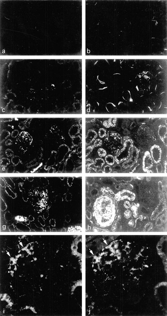
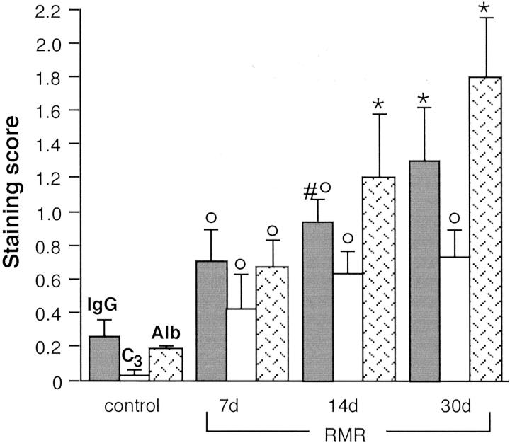
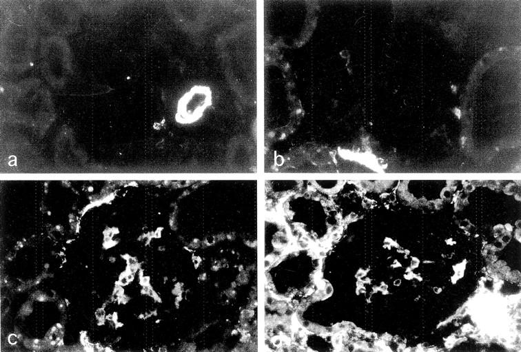
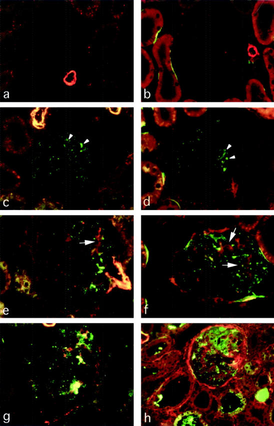
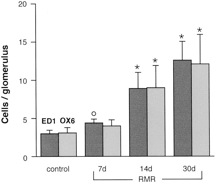
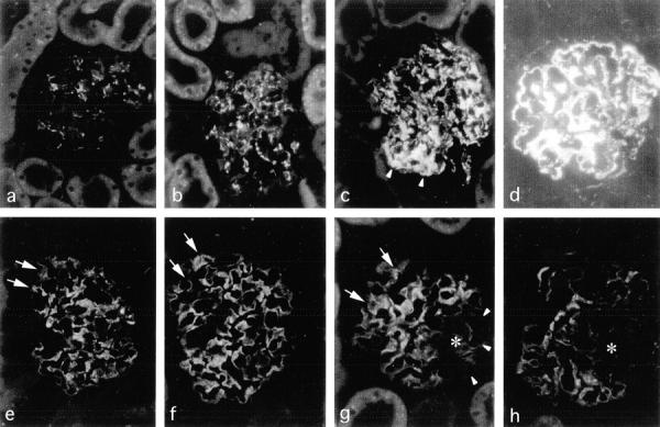
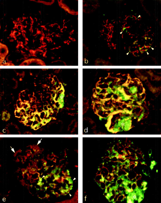
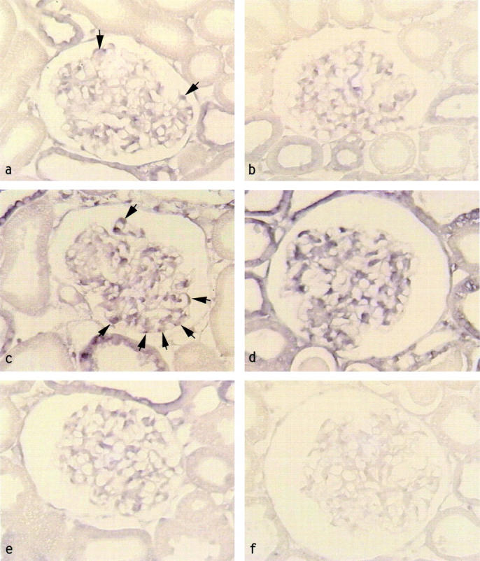
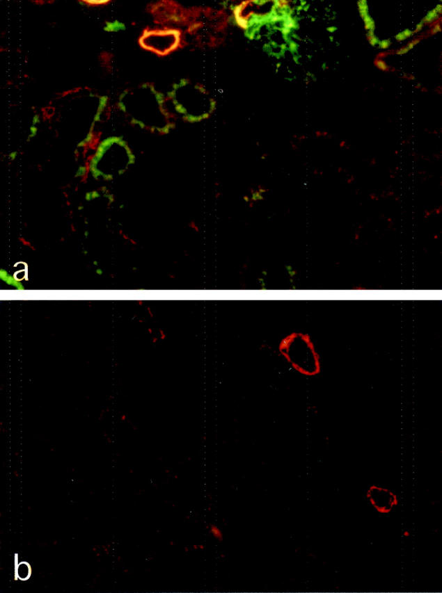
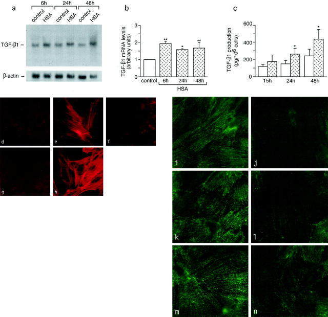
Chronic diseases of the kidney have a progressive course toward organ failure. Common pathway mechanisms of progressive injury, irrespectively of the etiology of the underlying diseases, include glomerular capillary hypertension and enhanced passage of plasma proteins across the glomerular capillary barrier because of impaired permselective function. These changes are associated with podocyte injury and glomerular sclerosis. Direct evidence for causal roles is lacking, particularly for the link between intraglomerular protein deposition and sclerosing reaction. Because transforming growth factor-beta1 (TGF-beta1) is the putative central mediator of scarring, we hypothesized that TGF-beta1 can be up-regulated by protein overload of podocytes thereby contributing to sclerosis. In rats with renal mass reduction, protein accumulation in podocytes as a consequence of enhanced transcapillary passage preceded podocyte dedifferentiation and injury, increase in TGF-beta1 expression in podocytes, and TGF-beta1-dependent activation of mesangial cells. Angiotensin-converting enzyme inhibitor prevented both accumulation of plasma proteins and TGF-beta1 overexpression in podocytes and sclerosis. Albumin load on podocytes in vitro caused loss of the synaptopodin differentiation marker and enhanced TGF-beta1 mRNA and protein. Conditioned medium of albumin-stimulated podocytes induced a sclerosing phenotype in mesangial cells, an effect mimicked by TGF-beta1 and blocked by anti-TGF-beta1 antibodies. Thus, the passage of excess plasma proteins across the glomerular capillary wall is the trigger of podocyte dysfunction and of a TGF-beta1-mediated mechanism underlying sclerosis. Agents to reduce TGF-beta1, possibly combined with angiotensin blockade, should have priority in novel approaches to treatment of progressive nephropathies.


Figures

Figure 1.
Figure 1.
Enhanced intraglomerular passage and accumulation of plasma proteins in RMR, a rat model of progressive nephropathy. a–h: Sections of kidneys after sham operation or RMR stained by immunofluorescence for IgG (left) or C3 (right). a and b, sham-operated control; c and d, RMR at 7 days, focal staining; e and f, RMR at 14 days, staining more evident both in glomeruli and in proximal tubuli; g and h, RMR at 30 days, heterogeneous patterns with strong irregular staining, or minimal amount of staining in one glomerulus in h. i and j: Abnormal albumin (i) and IgG staining (j) co-localize to the same podocytes (arrows) and mesangial areas of RMR kidney at 14 days. Note the glomerular intracellular staining of podocytes that reflects excess protein uptake. Direct immunofluorescence using FITC-conjugated antibodies. Original magnifications: ×125 (a–h); ×500 (i and j).
Figure 2.
Figure 2.
Semiquantitative analysis of IgG, C3, and albumin staining in glomeruli. °, P < 0.01 versus control; *, P < 0.01 versus control and RMR at 7 days; #, P < 0.05 versus 7 days RMR.
Figure 3.
Figure 3.
Expression of α-SMA in glomeruli in progressive nephropathy. a: Sham control, α-SMA staining confined to vessel wall and absent or minimal in glomeruli. b–d: RMR, b, at 7 days, absent or very weak α-SMA expression; c, at 14 days, strong focal staining and initial periglomerular expression; d, at 30 days, further increased expression in periglomerular tubulointerstitial areas. Original magnifications, ×250.
Figure 4.
Figure 4.
a–h: Double-immunofluorescence detection of α-SMA and IgG (left) or α-SMA and C3 (right) in RMR kidneys. α-SMA is stained in red (Cy-3-conjugated secondary antibody), IgG and C3 are stained in green (FITC-conjugated antibodies). a and b, sham control; c and d, RMR at 7 days; e and f, RMR at 14 days; g and h, RMR at 30 days. The comparison of α-SMA expression with both IgG and C3 deposition shows that the abnormal accumulation of plasma proteins (arrowheads) is detected both in advance of and in close association with the mesangial (arrows) and periglomerular myofibroblast transformation. Original magnifications, ×250.
Figure 5.
Figure 5.
Numbers of ED1 macrophages and MHC class II-positive cells in glomeruli. °, P < 0.01 versus control; *, P < 0.01 versus control and RMR at 7 days.
Figure 6.
Figure 6.
Immunofluorescence detection of desmin (a–d) and synaptopodin (e–h) in RMR glomeruli. In contrast to normal patterns revealed in sham control (a and e) and RMR at 7 days (b and f), high desmin expression and loss of synaptopodin in podocytes are visualized in RMR at 14 days (c and g, arrowheads) and 30 days (d and h) after surgery. In respect to areas in which synaptopodin is preserved (arrows), the asterisks indicate well-defined areas in which the foot process-associated differentiation marker is lost. Original magnifications, ×250.
Figure 7.
Figure 7.
Comparison of the glomerular sites of abnormal deposition of protein and altered expression of desmin and synaptopodin. a–d: Dual labeling of desmin and IgG. In a glomerulus of sham control (a) desmin (red) is confined to mesangial cells and IgG (green) is not detectable. A glomerulus of RMR kidney at 7 days (b) shows granular IgG staining (arrowheads) in podocytes, associated with no or very weak desmin staining, consistent with IgG accumulation in advance of high desmin expression. In sections of RMR kidneys at 14 days (c) and at 30 days (d) both the high expression of desmin and the IgG staining co-localize mainly to peripheral podocytes (yellow). e and f: By dual labeling for synaptopodin and IgG in RMR at 14 days (e) and at 30 (f), IgG-positive podocytes (arrowheads), in contrast to IgG-negative podocytes (arrows), show diminution or loss of synaptopodin (red). Severe podocyte damage and possibly detachment and loss are detectable in association with extracellular IgG deposition in areas of segmental adhesion and sclerosis (asterisks). Original magnifications, ×250.
Figure 8.
Figure 8.
Glomerular TGF-β1 up-regulation in RMR and effect of ACE inhibitor. TGF-β1 mRNA expression by in situ hybridization in glomeruli of a, sham control revealing weak signal in podocytes, b, RMR at 7 days; c, RMR at 14 days; d, RMR at 30 days; e, RMR at 30 days plus ACE inhibitor. TGF-β1 mRNA signal is increased in RMR at 14 days (c) in podocytes (arrows) and intracapillary areas, and it becomes more diffuse at 30 days (d). The ACE inhibitor prevents TGF-β1 up-regulation (e). f, RMR at 30 days, sense probe. Original magnifications, ×375.
Figure 9.
Figure 9.
Sections of RMR kidney at 30 days and RMR kidney of an ACEi-treated rat, stained for IgG (green) and α-SMA (red). Compared to untreated RMR (a), the ACE inhibitor prevents abnormal accumulation of IgG (b). Original magnifications, ×125.
Figure 10.
Figure 10.
Synthesis of TGF-β1, induction of downstream phenotypic change of mesangial cells, and loss of synaptopodin by podocytes in response to protein load. a–c: Effects of HSA on TGF-β1 mRNA expression by Northern blot analysis (a and b) and on TGF-β1 production by enzyme-linked immunosorbent assay (c) in cultured podocytes. *, P < 0.05 and **, P < 0.01 versus control. d–h: Expression of α-SMA in mesangial cells exposed to conditioned medium of unstimulated podocytes (d) or of HSA-stimulated podocytes alone (e) or in the presence of anti-TGF-β1 antibody (f), as assessed by fluorescence confocal microscopy. g and h: α-SMA in control mesangial cells either unstimulated (g) or exposed to TGF-β1 (h). i–n: Confocal microscopy analysis of the effect of HSA on podocyte synaptopodin. Podocytes exhibit marked reduction of synaptopodin staining after exposure to HSA for 6 hours (i) and 24 hours (l), and partial recovery at 48 hours (n) as compared to unstimulated control at the corresponding time points (i, k, and m). Original magnifications, ×1500.

References

    1. Brenner BM, Meyer TW, Hostetter TH: Dietary protein intake and the progressive nature of kidney disease: the role of hemodynamically mediated glomerular injury in the pathogenesis of progressive glomerular sclerosis in aging, renal ablation, and intrinsic renal disease. N Engl J Med 1982, 307:652-659 - PubMed
    1. Anderson S, Meyer TW, Rennke HG, Brenner BM: Control of glomerular hypertension limits glomerular injury in rats with reduced renal mass. J Clin Invest 1985, 76:612-619 - PMC - PubMed
    1. Fries JWU, Sandstrom DJ, Meyer TW, Rennke HG: Glomerular hypertrophy and epithelial cell injury modulate progressive glomerulosclerosis in the rat. Lab Invest 1989, 60:205-218 - PubMed
    1. Remuzzi G, Bertani T: Is glomerulosclerosis a consequence of altered glomerular permeability to macromolecules? Kidney Int 1990, 38:384-394 - PubMed
    1. Olson JL, Hostetter TH, Rennke HG, Brenner BM, Venkatachalam MA: Altered glomerular permselectivity and progressive sclerosis following extreme ablation of renal mass. Kidney Int 1982, 22:112-126 - PubMed

MeSH terms

LinkOut - more resources