. 2003 Jan;130(2):379-89.
doi: 10.1242/dev.00201.

'Cyclic alopecia' in Msx2 mutants: defects in hair cycling and hair shaft differentiation

Affiliations

'Cyclic alopecia' in Msx2 mutants: defects in hair cycling and hair shaft differentiation

Liang Ma et al. Development. 2003 Jan.

Abstract

Msx2-deficient mice exhibit progressive hair loss, starting at P14 and followed by successive cycles of wavelike regrowth and loss. During the hair cycle, Msx2 deficiency shortens anagen phase, but prolongs catagen and telogen. Msx2-deficient hair shafts are structurally abnormal. Molecular analyses suggest a Bmp4/Bmp2/Msx2/Foxn1 acidic hair keratin pathway is involved. These structurally abnormal hairs are easily dislodged in catagen implying a precocious exogen. Deficiency in Msx2 helps to reveal the distinctive skin domains on the same mouse. Each domain cycles asynchronously - although hairs within each skin domain cycle in synchronized waves. Thus, the combinatorial defects in hair cycling and differentiation, together with concealed skin domains, account for the cyclic alopecia phenotype.


Figures
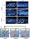
Fig. 1
Fig. 1
Expression of Msx1 and Msx2. Wild-type ICR pups were sacrificed at indicated time-points and their skin harvested, sectioned and hybridized to 35S-labeled Msx1 or Msx2 cRNA probes. Msx1 and Msx2 are initially co-expressed in the developing hair follicle. Expression of Msx1 is limited to the matrix cells (A,C) and its expression disappears during catagen (E). Msx2 expression in the hair follicle is more dynamic: first it is expressed only in the matrix and precortical cells, then it is expanded into the hair cortex and medulla (B,D). Msx2 continues to be expressed during catagen, when Msx1 expression is no longer expressed (E,F). Scale bar: 50 µm. (G) A schematic diagram of hair follicle structures and the hair cycle. During anagen, germinative matrix cells proliferate to generate progenitor cells, which receive signals from the dermal papilla to differentiate into either hair shaft cells or inner root sheath cells. During catagen, hair production ceases and hair follicle degenerates to form a club hair. During telogen, hair follicle rests and at the end of telogen, dermal papilla at the base of the hair follicle interact with the adjacent bulge region to initiate the second round of hair follicle morphogenesis. IRS, inner root sheath; ORS, outer root sheath; HF, hair follicle.
Fig. 2
Fig. 2
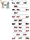
Abnormal hair cycling in Msx2 knockout mutant mouse. (A) Hair distribution of the same mouse, pictures taken 1 month apart. Note the dramatically different hair patches. In 1 month, hairy regions became bald, and bald regions became hairy. (B) Asynchronous hair cycle domains in the same mutant mouse over time. Msx2 knockout mice show a cyclic balding pattern. To examine the balding pattern, we followed five mice (1 month old) over an 80-day period, taking photos about once every 3 days. One example is shown here. Hair regrowth first appears in the shoulder region, spreads to the whole trunk, and then starts to get lost from cephalic to caudal end until all hairs are lost. This pattern then repeats. The times when most trunk hairs are in anagen are boxed in red.
Fig. 3
Fig. 3
Histopathological changes of Msx2 knockout mice in different hair cycle stages. Left column, normal cycling showed shortened anagen. Differences in skin and hair follicle morphology between wild-type and Msx2 knockout mice start to show at postnatal day 3 (P3). There is a lack of hair cortex differentiation at P5 (A,B). Anagen in Msx2 knockout mutant is shorter and hair follicles at P10 have already entered catagen (C,D). Catagen progression is also delayed compared with wild type. At P21, wild-type follicles have entered telogen, whereas Msx2 knockout mutant follicles are still in catagen (E,F). At P24, control skin has entered anagen, while Msx2 knockout mutants are still in telogen (G,H). Msx2 knockout mutant skin eventually re-entered anagen at P31. Examination of Tgfa expression supported the histological observation (E,F, insets). No Tgfa expression was detected in wild type hair follicles at P21 whereas strong Tgfa expression was still present in Msx2 knockout mutant hair follicles (arrow, in inset of F). Scale bars: 500 µm. Quantification (below) was carried out by counting about 50 hair follicles (at designated days after birth) and converting them into percentage of hair follicles in different hair cycle stages. Orange is anagen, green is catagen and blue is telogen. Error bar represents one standard deviation. (Right column) Regeneration after plucking showed defect in re-entry into anagen. One-month-old mice were stripped with hot wax and followed at 6, 10, 14 and 20 day post-stripping. Note that normal hair follicles re-enter anagen at day 6 (C,E), while those of Msx2 knockout mutants remain in telogen (D,F), and do not enter anagen until 20 days after stripping (H). At this time, control skin has re-entered telogen (G). Quantification is achieved as described in the development column. Scale bar: 500µm. Arrowheads in E,F indicate dermal papilla stained with alkaline phosphatase.
Fig. 4
Fig. 4
Cross between Msx2 and Fgf 5go/Fgf5go (Angora) mice. To address the genetic relationship between Msx2 and Fgf5 in hair cycle regulation, double mutant mice for Msx2 and Fgf5 were generated by crossing the two mutants. Hair loss in Msx2 knockout mice occurs invariably at P14 (A,B). By contrast, Fgf5go/Fgf5go mice grow long pelage hairs as a result of prolonged anagen (A). Mice doubly homozygous for both mutations exhibited long pelage hairs and no longer lose their pelage hairs at P14. Instead, hair loss in these mice eventually occurs between P18 and P30, depending on the genetic background of the mouse (B). (C) The hair cycle length in each genetic mutant. Anagen in double mutant is prolonged, similar to Fgf 5 mutants. Hair loss still occurs, but is also delayed accordingly. Catagen and telogen in double mutants are approximately similar to that of Msx2 mutants. The result suggests that hair loss in Msx2 knockout mutants is associated with a specific time-point in catagen and is delayed by mutation in Fgf5, which prolongs anagen length.
Fig. 5
Fig. 5
Changes of hair filament differentiation in Msx2 knockout mice. (A,B) Plucked hairs from Msx2 knockout mice are short and curly, reflecting defects in hair structures. Scale bar: 100 µm. (C,D) Medulla patterning is affected as suggested by the irregular septations in the hair. Scale bar: 50 µm. (E–H) Scanning EM showing unevenness in diameter of Msx2 knockout mice hairs. Cuticles fail to form, resulting in a smooth, wrinkled surface. Scale bar: 30 µm in E,F; 10 µm in G,H. (I,J) Trunk hairs from wild-type (I) and Msx2 knockout (J) mice. Both club ends are morphologically similar (arrowheads). Septation patterns are irregular in the mutants, but there are no breakages in the middle of the shaft. Scale bar: 100 µm. (K,L) BrdU labeling was performed at P5, P9, P11 and P15, and representative panels from P11 are shown. BrdU-positive cells were detected in both the hair matrix and the outer root sheath (arrows). Quantitation of BrdU-positive cells in the matrix showed similar levels of incorporation in the mutants. (M,N) Apoptotic cells detected by TUNEL assay carried out at P5, P9, P11, P14 and P30 (an example is shown from P30). Positive cells were observed in epidermis and dermis. No apparent differences between wild-type and Msx2 knockout mutant skins were observed. Scale bars: 250 µm.
Fig. 6
Fig. 6
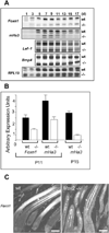
Changes of other molecular pathways in Msx2 knockout mutant hairs. (A) Timecourse RNase protection assays. Expression analyses of molecules implicated in hair differentiation. A 1 cm2 dorsal skin sample from indicated time points extracted for RNA. Examination of hair cortex differentiation markers by RNase protection assay revealed significantly lower levels of Foxn1 and its target gene Ha3 in Msx2 knockout mutants. At P13 (postnatal day; d on figure) Ha3 expression sharply decreases in the mutant skin and is barely detectable at P15 and P17 (A,C). This loss of Ha3 expression correlates with hair loss in Msx2 knockout mutants. Lef1 expression in the hair matrix cells and in the wild-type skin, increases from P7 to P11, which was not seen in Msx2 knockout mutants. Although the difference is not striking, the trend is consistent in different experiments. Expression of two other genes, Bmp4 and Tgfa (not shown) is not affected by the Msx2 mutation. Scale bar: 200 µm. (B) Quantitation of Foxn1 and Ha3 message levels at P11 revealed that Foxn1 and Ha3 mRNA is downregulated 50% compared with that in wild-type littermates. A much more dramatic 72% reduction in Ha3 expression was observed at P15. (C) Indirect immunohistochemistry with an affinity-purified Foxn1 antibody showed reduced Foxn1 protein in Msx2 knockout mutant cortex.
Fig. 7
Fig. 7
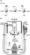
Summary diagram for hair shaft differentiation and hair cycle regulators. (A) Regulators and check points at each hair cycle phase transition. In essence, the hair cycle is orchestrated by molecules that regulate the transition between anagen-catagen, catagen-telogen and telogen-anagen. Each checkpoint is likely to be regulated by a group of factors. Some promote and some suppress the transition. The observed length of each hair cycle phase reflects the summation of the activities that promote or suppress the entry to the next phase. In the Msx2 knockout mutants, catagen starts earlier and lasts longer, and telogen hair has difficulty re-entering anagen. Therefore, it is most likely that the normal role of Msx2 in hair cycling is to maintain hairs in anagen phase. (B) Role of Msx2 in hair shaft differentiation. Upon induction of dermal papilla, stem cells in outer root sheath (ORS) generate TA cells that migrate to the matrix region. TA cells proliferate to generate cellular masses for making differentiated hair structures, and the regulation of this cellular flow can determine the size of hairs (Wang et al., 1999). The specified cell types are arranged in concentric layers from outside to inside. Several major molecular pathways are known to be involved in this specification and differentiation process (see text for detail). Msx2 is one of the central integrators that transmits growth factor signals to regulate hair differentiation.

Similar articles

Cited by

References

    1. Baxter RM, Brissette JL. Role of the nude gene in epithelial terminal differentiation. J. Invest. Dermatol. 2002;118:303–309. - PubMed
    1. Bechtold LS. Ultra-structural evaluation of mouse mutations. In: Sundberg JP, Boggess D, editors. Systematic Approach to Evaluation of Mouse Mutations. Boca Raton, FL: CRC Press; 2000. pp. 121–129.
    1. Botchkarev VA, Botchkareva NV, Roth W, Nakamura M, Chen LH, Herzog W, Lindner G, McMahon JA, Peters C, Lauster R, et al. Noggin is a mesenchymally derived stimulator of hair-follicle induction. Nat. Cell Biol. 1999;1:158–164. - PubMed
    1. Brissette JL, Li J, Kamimura J, Lee D, Dotto GP. The product of the mouse nude locus, Foxn1, regulates the balance between epithelial cell growth and differentiation. Genes Dev. 1996;10:2212–2221. - PubMed
    1. Chan EF, Gat U, McNiff JM, Fuchs E. A common human skin tumour is caused by activating mutations in beta-catenin. Nat. Genet. 1999;21:410–413. - PubMed

Publication types

MeSH terms