

Evidence for vestibular regulation of autonomic functions in a mouse genetic model
- PMID: 12466504
- PMCID: PMC139272
- DOI: 10.1073/pnas.252652299
Evidence for vestibular regulation of autonomic functions in a mouse genetic model
Abstract
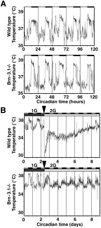
Physiological responses to changes in the gravitational field and body position, as well as symptoms of patients with anxiety-related disorders, have indicated an interrelationship between vestibular function and stress responses. However, the relative significance of cochlear and vestibular information in autonomic regulation remains unresolved because of the difficulties in distinguishing the relative contributions of other proprioceptive and interoceptive inputs, including vagal and somatic information. To investigate the role of cochlear and vestibular function in central and physiological responses, we have examined the effects of increased gravity in wild-type mice and mice lacking the POU homeodomain transcription factor Brn-3.1 (Brn-3bPou4f3). The only known phenotype of the Brn-3.1(-/-) mouse is related to hearing and balance functions, owing to the failure of cochlear and vestibular hair cells to differentiate properly. Here, we show that normal physiological responses to increased gravity (2G exposure), such as a dramatic drop in body temperature and concomitant circadian adjustment, were completely absent in Brn-3.1(-/-) mice. In line with the lack of autonomic responses, the massive increase in neuronal activity after 2G exposure normally detected in wild-type mice was virtually abolished in Brn-3.1(-/-) mice. Our results suggest that cochlear and vestibular hair cells are the primary regulators of autonomic responses to altered gravity and provide genetic evidence that these cells are sufficient to alter neural activity in regions involved in autonomic and neuroendocrine control.
Figures


Similar articles
-
Effects of a hair cell transcription factor, Brn-3.1, gene deletion on homozygous and heterozygous mouse cochleas in adulthood and aging.Hear Res. 1999 Aug;134(1-2):71-6. doi: 10.1016/s0378-5955(99)00070-2. Hear Res. 1999. PMID: 10452377
-
Essential role of POU-domain factor Brn-3c in auditory and vestibular hair cell development.Proc Natl Acad Sci U S A. 1997 Aug 19;94(17):9445-50. doi: 10.1073/pnas.94.17.9445. Proc Natl Acad Sci U S A. 1997. PMID: 9256502 Free PMC article.
-
Neurovestibular modulation of circadian and homeostatic regulation: vestibulohypothalamic connection?Proc Natl Acad Sci U S A. 2002 Nov 26;99(24):15723-8. doi: 10.1073/pnas.242251499. Epub 2002 Nov 14. Proc Natl Acad Sci U S A. 2002. PMID: 12434016 Free PMC article.
-
Vestibular autonomic regulation (including motion sickness and the mechanism of vomiting).Curr Opin Neurol. 1999 Feb;12(1):29-33. doi: 10.1097/00019052-199902000-00005. Curr Opin Neurol. 1999. PMID: 10097881 Review.
-
Vestibular autonomic regulation: overview and conclusions of a recent workshop at the University of Pittsburgh.J Vestib Res. 1998 Jan-Feb;8(1):1-5. J Vestib Res. 1998. PMID: 9416583 Review. No abstract available.
Cited by
-
Vestibular stimulation by 2G hypergravity modifies resynchronization in temperature rhythm in rats.Sci Rep. 2020 Jun 8;10(1):9216. doi: 10.1038/s41598-020-65496-x. Sci Rep. 2020. PMID: 32514078 Free PMC article.
-
A preformed scleral search coil for measuring mouse eye movements.J Neurosci Methods. 2010 Oct 30;193(1):126-31. doi: 10.1016/j.jneumeth.2010.08.023. Epub 2010 Sep 15. J Neurosci Methods. 2010. PMID: 20817027 Free PMC article.
-
Reviewing the Role of the Efferent Vestibular System in Motor and Vestibular Circuits.Front Physiol. 2017 Aug 2;8:552. doi: 10.3389/fphys.2017.00552. eCollection 2017. Front Physiol. 2017. PMID: 28824449 Free PMC article. Review.
-
The development of vestibular system and related functions in mammals: impact of gravity.Front Integr Neurosci. 2014 Feb 7;8:11. doi: 10.3389/fnint.2014.00011. eCollection 2014. Front Integr Neurosci. 2014. PMID: 24570658 Free PMC article. Review.
-
Efferent Vestibular Neurons Show Homogenous Discharge Output But Heterogeneous Synaptic Input Profile In Vitro.PLoS One. 2015 Sep 30;10(9):e0139548. doi: 10.1371/journal.pone.0139548. eCollection 2015. PLoS One. 2015. PMID: 26422206 Free PMC article.
References
-
- Ishihama L. M., Murakami, D. M. & Fuller, C. A. (1989) Physiologist 32, S61-S62. - PubMed
-
- Fuller P. M., Warden, C. H., Barry, S. J. & Fuller, C. A. (2000) J. Appl. Physiol. 89, 1491-1498. - PubMed
-
- Robinson E. L. & Fuller, C. A. (2000) Eur. J. Physiol. 441, R32-R38. - PubMed
-
- Pompeiano O., d'Ascanio, P., Centini, C., Pompeiano, M., Cirelli, C. & Tononi, G. (2001) Acta Otolaryngol. Suppl. 545, 120-126. - PubMed
Publication types
MeSH terms
Substances
Grants and funding
LinkOut - more resources
Full Text Sources
Other Literature Sources
Molecular Biology Databases
