. 2002 Dec 1;30(23):5017-28.
doi: 10.1093/nar/gkf656.

The 3' untranslated region of human vimentin mRNA interacts with protein complexes containing eEF-1gamma and HAX-1

Affiliations

The 3' untranslated region of human vimentin mRNA interacts with protein complexes containing eEF-1gamma and HAX-1

May Al-Maghrebi et al. Nucleic Acids Res. .

Abstract

Previously, we have shown that the vimentin 3' untranslated region (3'UTR) contains a highly conserved region, which is sufficient for the perinuclear localization of a reporter mRNA. This region was shown to specifically bind protein(s) by band shift analyses. UV-cross-linking studies suggest these proteins are 46- and 35-kDa in mass. Here, we have used this sequence as 'bait' to isolate RNA binding proteins using the yeast three-hybrid method. This technique relies on a functional assay detecting bona fide RNA-protein interaction in vivo. Three cDNA isolates, HAX-1, eEF-1gamma and hRIP, code for proteins of a size consistent with in vitro cross- linking studies. In all cases, recombinant proteins were capable of binding RNA in vitro. Although hRIP is thought to be a general mRNA binding protein, this represents an unreported activity for eEF-1gamma and HAX-1. Moreover, HAX-1 binding appears to be specific to vimentin's 3'UTR. Both in vivo synthesized eEF-1gamma and HAX-1 proteins were 'pulled out' of HeLa whole cell extracts by binding to a RNA affinity column comprised of vimentin's 3'UTR. Moreover, size-fractionation of extracts results in the separation of large complexes containing either eEF-1gamma or HAX-1. Thus, in addition to their known functions, both eEF-1gamma and HAX-1 are RNA binding proteins, which suggests new roles in mRNA translation and/or perinuclear localization.


Figures
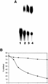
Figure 1
Figure 1
RNA band shift assay with HeLa WCE. (A) Twelve femtomoles of 32P-labeled RNA from the 3′UTR of human vimentin mRNA from position –37 to –123 downstream of the stop codon (37/123) was incubated alone (lane 1) or with increasing concentrations, 1 µg (lane 2), 2 µg (lane 3) or 4 µg (lane 4), of HeLa WCE. RNA–protein complexes were separated from free RNA on a 5% polyacrylamide gel, dried, and exposed to film overnight at –70°C. (B) 32P-labeled RNA as in (A) was incubated with 4 µg of HeLa WCE and increasing concentrations of unlabeled specific 37/123 (squares) and non-specific BMV (circles) competitor RNA. Data was quantified on a phosphoimage analyzer (Molecular Dynamics) and the percent of shifted material is plotted versus the fold-excess of cold RNA added.
Figure 2
Figure 2
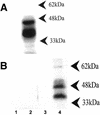
Analysis of RNA binding protein activity by UV cross-linking. (A) 32P-labeled RNA (37/147) (300 000 d.p.m.) was incubated with 20 µg of HeLa WCE. Following exposure to UV light at 254 nm for 15 min, non-cross-linked RNA was removed by digestion with RNase A (6 U) and T1 (1.5 U) for 2 h at 37°C. Proteins were separated on a 10% SDS–PAGE gel. The position of migration for several molecular weight markers is indicated by arrowheads. (B) Cross-linking studies were repeated in the presence of a 50-fold excess of non-specific γ-actin RNA (160/396) with 32P-labeled vimentin RNA (37/147) (lanes 2 and 4) or 32P-labeled γ-actin RNA (160/396) (lanes 1 and 3). HeLa WCE (20 µg) was incubated with the respective RNAs in lanes 2 and 4 whereas RNA alone was analyzed in lanes 1 and 3. The position of migration for several molecular weight markers is shown as in (A).
Figure 3
Figure 3
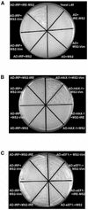
Yeast three-hybrid analysis of RNA–protein interaction monitored by activation of the HIS3 gene in vivo. Plasmids encoding the indicated hybrid RNAs [IRE-MS2, MS2-Vim (37/123) or MS2 alone] and AD fusions (AD-IRP, AD-Hax-1, AD-eEF-1γ or AD alone) were transformed into yeast strain L40 coat. After selecting for the presence of the plasmids, colonies were re-streaked onto media selecting for expression of HIS3. (A) Control plate showing that only the combination, which should lead to functional IRE/IRP interaction, yields growth in the presence of 5 mM 3-AT. (B) Experimental plate testing the interaction between AD-HAX-1 or AD-IRP fusions and various RNA samples grown on media containing 50 mM 3-AT. (C) Experimental plate testing the interaction between AD-eEF-1γ or AD-IRP and various RNA samples grown at 0.5 mM 3-AT.
Figure 4
Figure 4
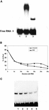
Analysis of the specificity of eEF-1γ binding by RNA band shift assays. (A) Band shift analysis as in Figure 1A with RNA alone (lane 1), 3 µg of HeLa WCE (lane 2) or 50 ng of purified recombinant eEF-1γ (lane 3). (B) Band shift analysis as in (A) (lane 3) with increasing concentrations of unlabeled specific RNA (diamonds) or non-specific, BMV RNA (triangles) as indicated. Data were quantified as discussed in the legend to Figure 1. (C) Band shift analysis as in (A) (lane 3) with 50 ng of recombinant eEF-1γ (lane 1) and increasing concentrations, 0.5, 2.5, 5 and 10 µģ, of anti-eEF-1γ in lanes 2–5, respectively.
Figure 5
Figure 5
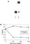
Analysis of the specificity of HAX-1 binding by RNA band shift assays. (A) Band shift analysis as in Figure 1A with RNA alone (lane 1) or 50 ng of purified recombinant HAX-1. (B) Band shift analysis as in (A) with increasing concentrations of specific (diamonds) or non-specific (circles) RNA competitor as noted. Data were quantified as discussed in the legend to Figure 1.
Figure 6
Figure 6
Western blot analysis of protein binding to the vimentin 3′UTR affinity column using anti-eEF-1γ. (A) Vimentin 3′UTR transcript (37/147) was annealed to 150 pmol of biotinylated oligonucleotide (complementary to the region 123/147) and immobilized on SA-PMP as described in Materials and Methods. Following incubation with HeLa WCE (277 μg) for 15 min at 4°C, the immobilized RNA transcript was washed twice with binding buffer (lanes 4 and 5, respectively), eluted with water (lane 1), and boiled with SDS-loading buffer followed by analysis of the supernatant (lane 2). All samples were analyzed on a 10% SDS–PAGE gel. For reference initial HeLa WCE extract (lane 6), purified recombinant eEF-1γ (lane 7) and protein, which flowed through the column (lane 3), was analyzed accordingly. (B) Identical to (A) except that the vimentin 3′UTR RNA transcript used was from the region 127/139 only, i.e., it did not contain the RNA–protein binding site. (C) Identical to (A) except that HeLa WCE was incubated with SA-PMP alone. There was no vimentin 3′UTR transcript or biotinylated oligonucleotide present. Additional details of the western blot protocol are described in Materials and Methods.
Figure 7
Figure 7
Western blot analysis of protein binding to the vimentin 3′UTR affinity column using anti-HAX-1. (AC) All panels same as in Figure 6, but analyzed with anti-HAX-1 antibody. For reference purified recombinant HAX-1 is loaded in lane 7.
Figure 8
Figure 8
Fractionation of RNA binding activity on a Superose 6 column. (A) HeLa cytoplasmic extract (30 mg) was fractionated on a Superose 6 column as described in Materials and Methods. Band shift activity (using 32P-labeled 37/147 RNA) was assessed for each fraction (10 µl) as well as the starting material (CE). Only those fractions displaying band shift activity (fraction nos 32–52) are shown. One band (*) was common to all fractions and thus, was deemed non-specific. An arrow denotes that band shift species which contains HAX-1 whereas an arrowhead identifies band shift activity due to eEF-1γ. The mobility of the free RNA is noted. Molecular weight standards of 200- and 45-kDa (K) eluted at fractions 37/38 and 45, respectively. (B) Western blotting analysis of each fraction (14 µl) probed with anti-HAX-1 antibody as described in Materials and Methods. Only those fractions shown to contain HAX-1 antigen are displayed. Cytoplasmic extract is analyzed in the lane marked CE. The position of migration of a 35-kDa standard (white band) is noted in the lane marked Std. (C) Western blotting analysis of each fraction (12 µl) probed with anti-eEF-1γ antibody. Only those fractions containing the eEF-1γ antigen are shown. The mobility of recombinant eEF-1γ (rEF) is noted. Fractions 37 and 39 were loaded in the outside lane of two different gels, hence the slight curvature in mobility.

Similar articles

Cited by

References

    1. Ross J. (1996) Control of mRNA stability in higher eukaryotes. Trends Genet., 12, 171–175. - PubMed
    1. Hunt T. (1989) On the translational control of suicide in red cell development. Trends Biochem. Sci., 14, 393–394. - PubMed
    1. Lindquist J.N., Kauschke,S.G., Stefanovic,B., Burchardt,E.R. and Brenner,D.A. (2000) Characterization of the interaction between αCP2 and the 3′-untranslated region of collagen α1(I) mRNA. Nucleic Acids Res., 28, 4306–4316. - PMC - PubMed
    1. Ostareck-Lederer A., Ostareck,D.H. and Hentze,M.W. (1998) Cytoplasmic regulatory functions of the KH-domain proteins hnRNPs K and E1/E2. Trends Biochem. Sci., 23, 409–411. - PubMed
    1. Wreden C., Verrotti,A.C., Schisa,J.A., Lieberfarb,M.E. and Strickland,S. (1997) Nanos and pumilio establish embryonic polarity in Drosophila by promoting posterior deadenylation of hunchback mRNA. Development, 124, 3015–3023. - PubMed

Publication types