. 2002 Dec 1;30(23):5065-73.
doi: 10.1093/nar/gkf670.

Elucidation of structure-function relationships in the protein subunit of bacterial RNase P using a genetic complementation approach

Affiliations

Elucidation of structure-function relationships in the protein subunit of bacterial RNase P using a genetic complementation approach

Milan Jovanovic et al. Nucleic Acids Res. .

Abstract

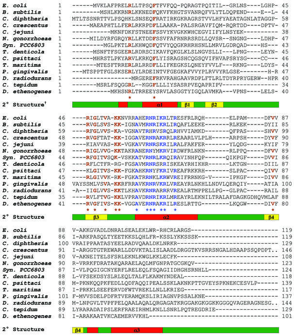
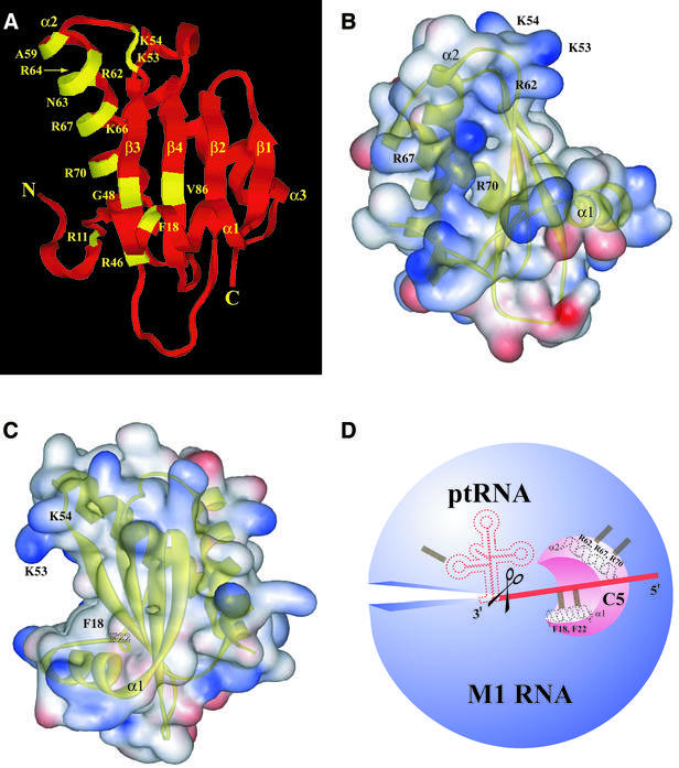
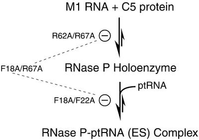
RNase P is a ribonucleoprotein involved in tRNA biosynthesis in all living organisms. Bacterial RNase P is comprised of a catalytic RNA subunit and a lone protein cofactor which plays a supporting, albeit essential, role in the tRNA processing reaction in vivo. In this study, we have searched various databases to identify homologs of the protein subunit of RNase P from diverse bacteria and used an alignment of their primary sequences to determine the most highly conserved residues, and thereby extend earlier predictions of which residues might play an important role in RNA recognition. By employing a genetic complementation assay, we have also gained insights into structure- function relationships in the protein subunit of bacterial RNase P.


Figures

Figure 1
Figure 1
Sequence alignment of the protein subunit of RNase P from various bacteria. Although a ClustalW-based alignment of 112 homologs from 10 different phylogenetic groups of Bacteria was performed, only 14 representative sequences (E.coli, B.subtilis, Corynebacterium diphtheriae, Caulobacter crescentus, Campylobacter jejuni, Neisseria gonorrhoeae, Synechocystis sp. PCC 6803, Treponema denticola, Chlamydophila psittaci, Thermotoga maritima, Porphyromonas gingivalis, Deinococcus radiodurans, Chlorobium tepidum and Dehalococcoides ethenogenes) are shown in this illustration. Two sets of conserved residues at positions which exhibit at least 67% identity in the complete alignment are highlighted in blue and brown. The demarcation of secondary structure elements is based on the tertiary structure of the protein subunit of B.subtilis RNase P (22).
Figure 2
Figure 2
(A) Location of conserved residues in the tertiary structure of the protein subunit of bacterial RNase P. Although the illustrations are based on the tertiary structure of the protein subunit of B.subtilis RNase P, the numbering is based on the E.coli counterpart. (B and C) Web Lab Viewer-generated views of the electrostatic surface potential map of the protein subunit of B.subtilis RNase P in which the RNR motif (B) and the putative ptRNA substrate-binding cleft (C) are depicted. Regions containing basic and acidic residues are depicted in blue and red, respectively. Labels for the residues coincide with the location of the side chains. (D) A schematic depicting RNA recognition sites in the bacterial RNase P holoenzyme. Potential interactions between (i) Arg62, Arg67 and Arg70 in helix α2 of C5 protein and M1 RNA; and (ii) Phe18 and Phe22 in helix α1 of C5 protein and the ptRNA leader sequence, are illustrated. The scissors indicate the site of cleavage on the ptRNA molecule by bacterial RNase P.
Figure 3
Figure 3
Results of the genetic complementation assay. The growth phenotypes observed at 30 and 43°C for various mutant derivatives of C5 protein are indicated. The key for the various mutant derivatives is provided in Table 3.
Figure 4
Figure 4
Western blot analysis to examine the levels of expression of various mutant derivatives of C5 protein in E.coli NHY322 cells. Lane M, magic markers/size standards (Invitrogen); lane 1, wild type C5 protein; lane 2, pBR322 (vector control); lane 3, C5 R46H/R62A; lane 4, C5 R46H/K66A; lane 5, C5 R46H/R67A; lane 6, C5 R46H/R70A; lane 7, C5 F18A/F22A; lane 8, C5 F22A/R62A; lane 9, C5 F22A/R67A; lane 10, C5 F18A/R62A; lane 11, C5 F18A/K66A; lane 12, C5 F18A/R67A; lane 13, C5 R62A/R67A; lane 14, C5 R62A/R70A; lane 15, C5 R67A/R70A; and lane 16, purified C5 protein (20 ng).
None
Scheme 1.

Similar articles

Cited by

References

    1. Altman S. and Kirsebom,L.A. (1999) Ribonuclease P. In Gesteland,R.F., Cech,T. and Atkins,J.F. (eds), The RNA World, 2nd Edn. Cold Spring Harbor Laboratory Press, Cold Spring Harbor, NY, pp. 351–380.
    1. Harris M.E., Frank,D. and Pace,N.R. (1998) Structure and catalytic function of the bacterial ribonuclease P ribozyme. In Simons,R.W. and Grunberg-Manago,M. (eds), RNA structure and function. Cold Spring Harbor Laboratory Press, Cold Spring Harbor, NY, pp. 309–337.
    1. Guerrier-Takada C., Gardiner,K., Marsh,T., Pace,N.R. and Altman,S. (1983) The RNA moiety of ribonuclease P is the catalytic subunit of the enzyme. Cell, 35, 849–857. - PubMed
    1. Pannucci J.A., Haas,E.S., Hall,T. and Brown,J.W. (1999) RNase P RNAs from Archaea are catalytically active. Proc. Natl Acad. Sci. USA, 96, 7803–7808. - PMC - PubMed
    1. Schedl P. and Primakoff,P. (1973) Mutants of Escherichia coli thermosensitive for the synthesis of transfer RNA. Proc. Natl Acad. Sci. USA, 70, 2091–2095. - PMC - PubMed

Publication types

MeSH terms