. 2003 Jan;9(1):87-92.
doi: 10.1038/nm807. Epub 2002 Dec 9.

Plasmodium falciparum erythrocyte invasion through glycophorin C and selection for Gerbich negativity in human populations

Affiliations

Plasmodium falciparum erythrocyte invasion through glycophorin C and selection for Gerbich negativity in human populations

Alexander G Maier et al. Nat Med. 2003 Jan.

Abstract

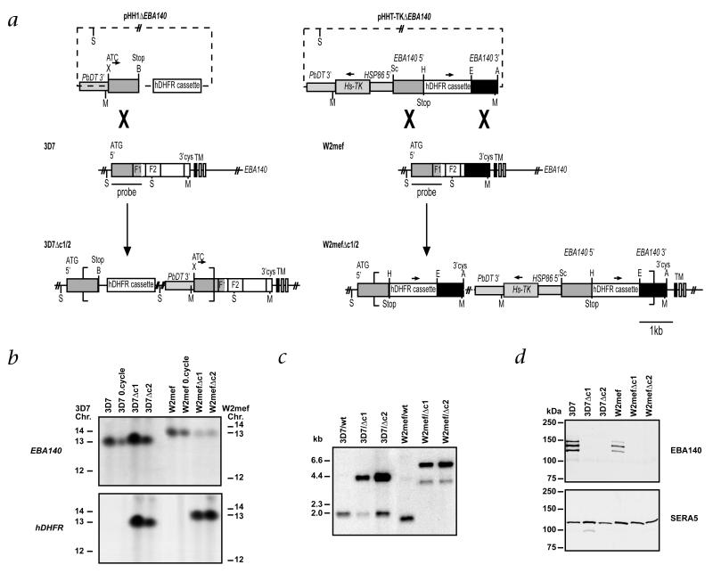
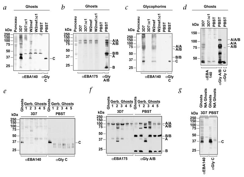
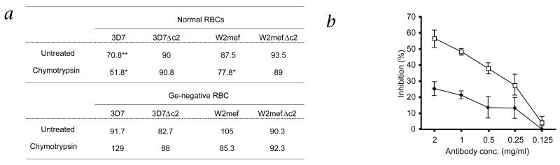
Geographic overlap between malaria and the occurrence of mutant hemoglobin and erythrocyte surface proteins has indicated that polymorphisms in human genes have been selected by severe malaria. Deletion of exon 3 in the glycophorin C gene (called GYPCDeltaex3 here) has been found in Melanesians; this alteration changes the serologic phenotype of the Gerbich (Ge) blood group system, resulting in Ge negativity. The GYPCDeltaex3 allele reaches a high frequency (46.5%) in coastal areas of Papua New Guinea where malaria is hyperendemic. The Plasmodium falciparum erythrocyte-binding antigen 140 (EBA140, also known as BAEBL) binds with high affinity to the surface of human erythrocytes. Here we show that the receptor for EBA140 is glycophorin C (GYPC) and that this interaction mediates a principal P. falciparum invasion pathway into human erythrocytes. EBA140 does not bind to GYPC in Ge-negative erythrocytes, nor can P. falciparum invade such cells using this invasion pathway. This provides compelling evidence that Ge negativity has arisen in Melanesian populations through natural selection by severe malaria.


Figures

Fig. 1
Fig. 1
Disruption of the gene encoding EBA140 in P. falciparum. a, Top, transfection plasmids pHH1ΔEBA140 and pHHT-TKΔEBA140 with selection cassette containing human DHFR and the Pb-DT3′ sequence; the pHHT-TKΔEBA140 plasmid contains the thymidine kinase cassette (Hs-TK). Middle, structure of the endogenous EBA140 gene for 3D7 and W2mef (F1 and F2 domains indicated). Bottom, integration into 3D7 EBA140 occurs by a single homologous recombination event; into W2mef, by a double homologous recombination event (more than one copy inserted so Hs-TK is retained). S, ScaI; Sc, SacII; B, BglII; X, XhoI; H, HpaI; E, EcoRI; A, AvrII; M, MfeI. b, Analysis of chromosomes from 3D7- and W2mef-transfected parasites. Chromosomes from 3D7 and W2mef; 3D7 and W2mef after one cycle of selection for integration (0.cycle); and cloned lines 3D7Δc1, 3D7Δc2 W2mefΔc1 and W2mefΔc2 were separated by pulsed-field gel electrophoresis and probed with genes encoding EBA140 or human DHFR (hDHFR). The EBA140 probe hybridizes to chromosome 13 in 3D7 and W2mef as well as in the transfected lines, indicating integration into this chromosome. This is confirmed by hybridization of the hDHFR probe, which detects chromosome 13 in the transfected cloned lines 3D7Δc1, 3D7Δc2 and W2mefΔc1 and W2mefΔc2. The ‘0.cycle’ parasites also hybridizes to the hDHFR probe, but to episomal plasmid that ran off the gel here. Left and right margins, chromosomal positions (Chr.). c, Southern blot analysis of genomic DNA confirming disruption of gene encoding EBA140 in 3D7 and W2mef. Genomic DNA from 3D7, Wmef and the cloned transfected lines 3D7Δc1, 3D7Δc2, W2mefΔc1 and W2mefΔc2 was digested with MfeI and ScaI and hybridized with the 5′ region of the gene encoding EBA140. d, Western blot analysis of supernatants from 3D7, W2mef and the transfected clones 3D7Δc1, 3D7Δc2, W2mefΔc1 and W2mefΔc2, using antibodies against EBA140. No bands are seen in the transfected clones, confirming that disruption of the gene encoding EBA140 results in loss of expression of EBA140. SERA5, another secreted protein (loading control). Left margins (c and d), molecular size markers.
Fig. 2
Fig. 2
EBA140 binds to GYPC on human erythrocytes. a, EBA140 binds to GYPC and other proteins in erythrocyte ghost preparations. Normal erythrocyte ghost cells were separated by SDS–PAGE, blotted and incubated with merozoite supernatants from 3D7, 3D7Δc1, W2mef and W2mefΔc1 (above blots). Far left lane, Ponceau staining of total erythrocytes proteins. PBST (far right lanes), ghost erythrocytes probed with antibodies against EBA140 or GYPC. b, EBA175 binds to GYPA homodimers and GYPA–GYPB heterodimers. Erythrocyte ghost proteins were incubated with supernatants from 3D7, W2mef, 3D7Δc1 and W2mefΔc1. Far left lane, Ponceau staining. PBST (far right lanes), probed directly with antibodies against EBA175 or GYPA–GYPB without prior incubation with supernatant. c, EBA140 binds to purified glycophorin proteins. Purified glycophorins separated by SDS–PAGE were incubated with supernatants from 3D7, W2mef, 3D7Δc1 and W2mefΔc1. Far left lane, Ponceau staining. PBST (far right lanes), probed with antibodies against EBA140 or GYPC. Right, GYPB and GYPC monomers. The GYPA dimer migrates at 75 kDa here. This glycophorin preparation consists mainly of GYPA but also has some GYPB and small amounts of GYPC. d, EBA140 binds to proteins the same size as GYPA and GYPA/B dimers. Samples similar to those in a and b were separated by 6% acrylamide gel electrophoresis and incubated with supernatants (above gel) to more distinctly separate the larger-molecular-weight bands to which EBA140 binds. PBST (far right lanes), probed with antibodies against EBA140, GYPA/GYPB or GYPC. e, EBA140 does not bind to mutant GYPC. Proteins from ghost erythrocytes obtained from normal or Ge-negative individuals (GYPCΔex3 homozygotes) were incubated with supernatant from 3D7 (above blot). Binding of EBA140 was detected with antibodies against EBA140. Left, an identical membrane probed with antibodies against GYPC. f, EBA175 binds to GYPA dimers in Ge-negative erythrocytes. A membrane identical to that in e was incubated with supernatant from 3D7 followed by detection of bound EBA175 with antibodies against EBA175. Right, incubation with antibodies against GYPA–GYPB. Ge-negative erythrocytes (lane 2) also have a mutant GYPB. g, Binding of EBA140 to erythrocytes is sialic acid-dependent. Proteins from normal ghost erythrocytes or those treated with neuraminidase (NA) were incubated with supernatants from 3D7 (left) and bound EBA140 identified with antibodies against EBA140. PBST (far right lanes), GYPC detected using antibodies against GYPC. Left margins, molecular size markers. Right margins, A/A, GYPA homodimer; A/B, GYPA/B heterodimer; B/B, GYPB homodimer; A, GYPA monomer; B, GYPD monomer; C, GYPC. α, antibody against; Gly, glycophorin.
Fig. 3
Fig. 3
Antibodies against the EBA140 F2 domain inhibit the EBA140–GYPC invasion pathway of P. falciparum. a, Data represent percentage of parasite invasion in the presence of EBA140 F2 antibodies relative to invasion in the presence of nonspecific antibodies from pre-immune serum. The inhibition of 3D7 wild-type parasites is more profound when red blood cells (RBCs) are treated with chymotrypsin. Chymotrypsin will cleave GYPB but not GYPC; this limits the receptor repertoire on the surface of the chymotrypsin-treated erythrocytes, enabling the dissection of invasion through GYPC versus other receptors. Conversely, inhibition by antibody against EBA140 is abolished when invasion of ΔEBA140 parasites is tested because of the lack of expression of EBA140. No inhibition is detectable when Ge-negative RBCs are used because they lack a GYPC receptor capable of binding to EBA140. Data represent the average of at least three experiments done in triplicate. *, P < 0.05 and **, P < 0.01, wild-type compared with EBA140-null parasite lines (paired t-test). b, Inhibition of invasion of 3D7 wild-type parasites by the EBA140 F2 antibody is concentration dependent. Data represent percent of inhibition of 3D7 wild-type invasion (◆) after addition of antibody against EBA140 F2 compared with that in the presence of antibodies of pre-immune serum (antibody concentration, horizontal axis). The inhibitory effect of the EBA140 F2 antibody on invasion is more profound for erythrocytes pre-treated with chymotrypsin (□). Error bars indicate confidence levels (α = 0.1) of five independent experiments done in triplicate.

References

    1. Haldane JBS. The rate of mutation in human genes. In: bonnier GA, editor. Proceedings of the VII International Congress on Genetics. 1949. pp. 267–273.
    1. Miller LH, Good MF, Milon G. Malaria pathogenesis. Science. 1994;264:1878–1883. - PubMed
    1. Booth PB, McLoughlin K. The Gerbich blood group system, especially in Melanesians. Vox Sang. 1972;22:73–84. - PubMed
    1. Serjeantson SW, White BS, Bhatia K, Trent RJ. A 3.5 kb deletion in the gly-cophorin C gene accounts for the Gerbich-negative blood group in Melanesians. Immunol. Cell Biol. 1994;72:23–27. - PubMed
    1. Patel SS, et al. The association of the glycophorin C exon 3 deletion with ovalocytosis and malaria susceptibility in the Wosera, Papua New Guinea. Blood. 2001;98:3489–3491. - PubMed

Publication types

MeSH terms