Review
. 2003:65:851-79.
doi: 10.1146/annurev.physiol.65.092101.142611. Epub 2002 May 1.

G protein-coupled receptor rhodopsin: a prospectus

Affiliations
Review

G protein-coupled receptor rhodopsin: a prospectus

Sławomir Filipek et al. Annu Rev Physiol. 2003.

Abstract

Rhodopsin is a retinal photoreceptor protein of bipartite structure consisting of the transmembrane protein opsin and a light-sensitive chromophore 11-cis-retinal, linked to opsin via a protonated Schiff base. Studies on rhodopsin have unveiled many structural and functional features that are common to a large and pharmacologically important group of proteins from the G protein-coupled receptor (GPCR) superfamily, of which rhodopsin is the best-studied member. In this work, we focus on structural features of rhodopsin as revealed by many biochemical and structural investigations. In particular, the high-resolution structure of bovine rhodopsin provides a template for understanding how GPCRs work. We describe the sensitivity and complexity of rhodopsin that lead to its important role in vision.


Figures

Figure 1
Figure 1
Molecular packing of rhodopsin (A) and sensory rhodopsin (B) in their crystals. One asymmetric unit contains two rhodopsin molecules. The central dimer is shown as a continuous surface, whereas the rest are shown as ribbons. Views are from different crystallographic X-, Y-, Z-axes for rhodopsin (27), and X-, and Z- for sensory rhodopsin. The data for sensory rhodopsin were taken from Protein Data Base (1H68). Coordinates: Models of rhodopsin and the homology models of cone pigments have been deposited in the Protein Data Bank (PDB) (identifiers 1F88, 1HZX, 1KPN, 1KPW, 1KPX).
Figure 2
Figure 2
Ribbon drawings of rhodopsin. Helices I–VIII are colored as a spectrum of visible light from blue (helix I) to red (helix VIII), and two orientations are shown. Palmitoyl chains and oligosaccharide groups shown using ball-and-stick models.
Figure 3
Figure 3
Two-dimensional model of bovine rhodopsin. Buried residues are shown in gray. Orange, red, and violet denote surface residues. Residues in contact with polar headgroups of the membrane on the cytoplasmic side are orange. Similar residues on the extracellular side are red. All other surface residues are violet.
Figure 4
Figure 4
Hydrophobic and hydrophilic interactions between rhodopsin helices (loops not included) viewed from the intracellular side. Hydrophobic interactions are shown as orange fuzzy bars together with numbers denoting their strength. Hydrophilic interactions are shown as red lines. List of hydrophilic interactions between helices include helices I–II, Asn55-Asp83; helices I–VII, Tyr43-Phe293, Asn55-Ala299; helices I–VIII, Gln64-Thr320; helices II–III, Asn78-Ser127; helices II–IV: Tyr74-Glu150, Asn78-Thr160, Asn78-Trp161; helices III–V, Cys140-Thr229, Glu122-Trp126-His211-Tyr206 network; helices III–VI, Arg135-Glu247; helices III–VII, Glu113-Lys296 (retinal); helices IV–V, Ala166-Tyr206; helices VI–VII, Cys264-Thr297; and helices VII–VIII, Ile307-Arg314.
Figure 5
Figure 5
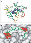
Vicinity of retinal in the binding site of rhodopsin. (A) Chromophore-binding site of bovine rhodopsin. The chromophore is in purple, and colors of helices are as in Figure 2. (B) The structure of 11-cis-retinal bovine opsin (27) using space-filling model. In blue are nitrogen atoms of the peptide bond and the Schiff base, with the hydrogen between Lys296 and the retinal in green. In red, two acidic residues in the binding site, Glu113 and Glu122, which is close to the β-ionone ring.
Figure 6
Figure 6
The DRY region (A) and NPXXY region (B) are believed to be involved in the activation and transformation of photoactivated rhodopsin. Note that in rhodopsin, the sequence of the DRY region is ERY.
Figure 7
Figure 7
Interactions of helix VI. Hydrophobic contacts of helix VI shown as white residues and hydrophilic interactions represented by lines. (A) Interaction of helix VI with helices II and III. (B) Interaction of helix VI with helices V and VII.
Figure 8
Figure 8
Different regions of cone pigments. (A) DRY region and the chromophore-binding sites of blue and red/green cone opsins. (B) The vicinity of the chromophore from left to right: blue, green, and red pigments. The numbering of corresponding regions is based on the bovine rhodopsin primary sequence.
Figure 9
Figure 9
Functional domains of rhodopsin that are highly conserved among members of the GPCR superfamily. (A) Location of these domains in the three-dimensional structure of rhodopsin. (B) Close-up of the critical regions: DRY region (a, panel A, in rhodopsin ERY, blue); palmitoyl groups (b, panel A, red); NPXXY region (c, panel A, light blue); chromophore; Pro kink in helix VI, Lys296 (d, panel A, violet); disulfide bridge (e, panel A, yellow); and oligosaccharide moieties (f, panel A, brown).

References

    1. Kühne W. Ueber den Sehpurpur. Unters. Physiol. Inst. Univ. Heidelberg; Heidelberg: 1878. p. 113.
    1. Wald G. Carotenoids and the visual cycle. J. Gen. Physiol. 1935–1936;19:351–71. - PMC - PubMed
    1. Hubbard R, Kropf A. Molecular isomers in vision. Sci. Am. 1967;216:64–70. - PubMed
    1. Wald G. The molecular basis of visual excitation. Nature. 1968;219:800–7. - PubMed
    1. Matthews RG, Hubbard R, Brown PK, Wald G. Tautomeric forms of meta-rhodopsin. J. Gen. Physiol. 1963;47:215–40. - PMC - PubMed

Publication types

LinkOut - more resources