. 2002 Dec 9;159(5):783-93.
doi: 10.1083/jcb.200204149. Epub 2002 Dec 9.

Lamin A/C speckles mediate spatial organization of splicing factor compartments and RNA polymerase II transcription

Affiliations

Lamin A/C speckles mediate spatial organization of splicing factor compartments and RNA polymerase II transcription

R Ileng Kumaran et al. J Cell Biol. .

Abstract

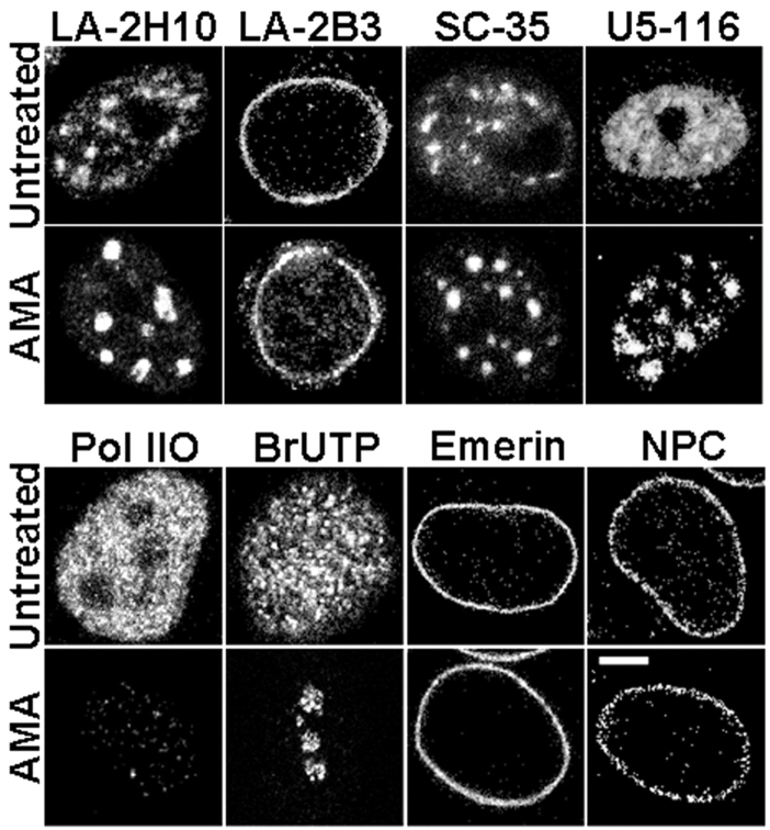
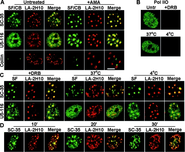
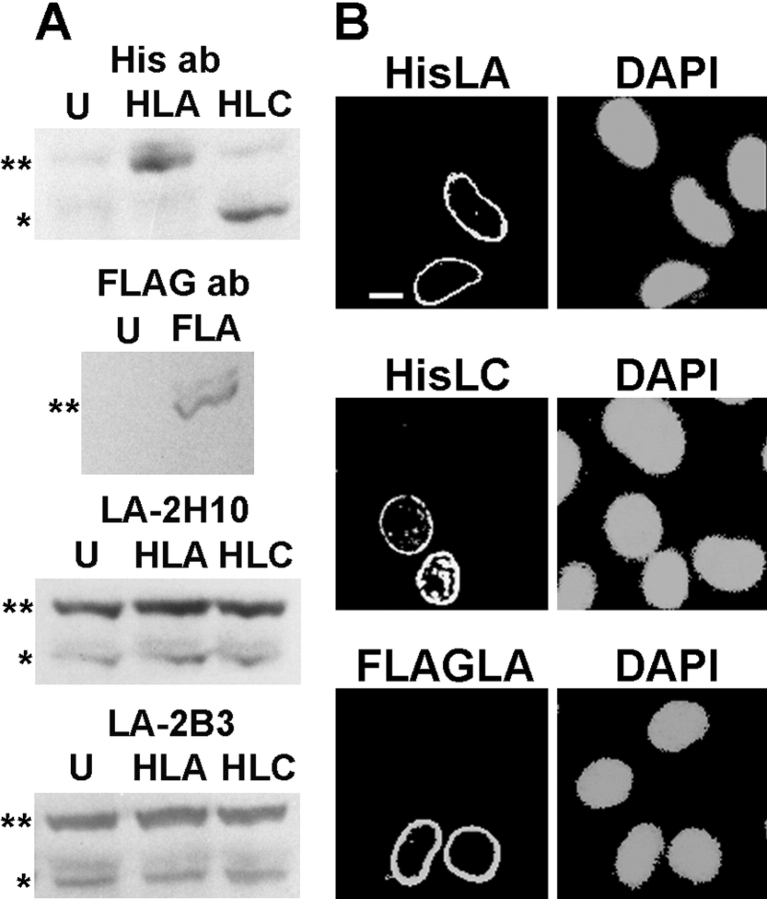
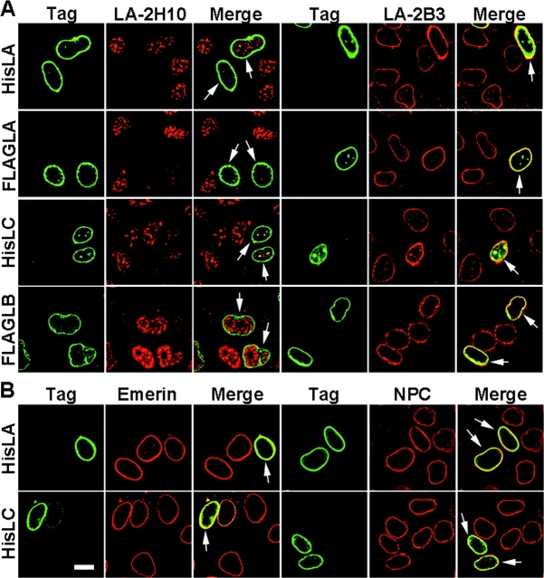
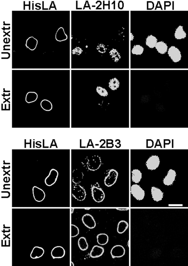
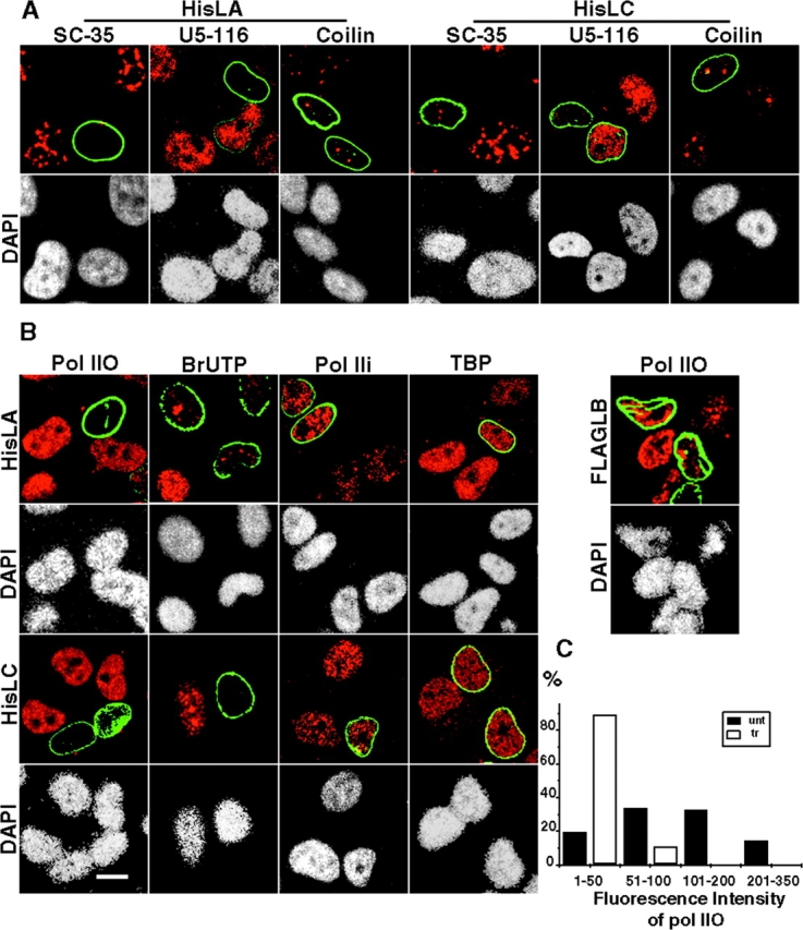
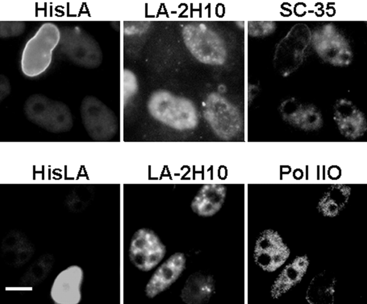
The A-type lamins have been observed to colocalize with RNA splicing factors in speckles within the nucleus, in addition to their typical distribution at the nuclear periphery. To understand the functions of lamin speckles, the effects of transcriptional inhibitors known to modify RNA splicing factor compartments (SFCs) were examined. Treatment of HeLa cells with alpha-amanitin or 5,6-dichlorobenzimidazole riboside (DRB) inhibited RNA polymerase II (pol II) transcription and led to the enlargement of lamin speckles as well as SFCs. Removal of the reversible inhibitor DRB resulted in the reactivation of transcription and a rapid, synchronous redistribution of lamins and splicing factors to normal-sized speckles, indicating a close association between lamin speckles and SFCs. Conversely, the expression of NH2-terminally modified lamin A or C in HeLa cells brought about a loss of lamin speckles, depletion of SFCs, and down-regulation of pol II transcription without affecting the peripheral lamina. Our results suggest a unique role for lamin speckles in the spatial organization of RNA splicing factors and pol II transcription in the nucleus.


Figures

Figure 1.
Figure 1.
Lamin speckles form enlarged foci upon α-amanitin treatment. HeLa cells were treated with α-amanitin (AMA), fixed in formaldehyde, and labeled with antibodies to lamin A (LA-2H10 and LA-2B3), RNA splicing factors (SC-35 and U5-116 kD), RNA pol IIO, BrUTP (after RNA synthesis assay), emerin, and NPC proteins. Bar, 5 μm.
Figure 2.
Figure 2.
Localization of lamin speckles and SFCs in enlarged foci upon treatment with transcriptional inhibitors, and their synchronous redistribution after removal of DRB. (A) HeLa cells treated with α-amanitin (AMA) were fixed in formaldehyde and doubly labeled with mAb LA-2H10 and antibodies to splicing factors or Cajal bodies (SF/CB) as indicated. (B and C) HeLa cells were treated with DRB, washed, incubated for 1 h at 37°C or 4°C in complete DME, formaldehyde fixed, and labeled with antibodies to pol IIO alone (B) or splicing factors and LA-2H10 (double labeling) (C). (D) Time course of redistribution of lamin speckles and SC-35 after DRB treatment, washing, and incubation at 37°C for 10–30 min. Single optical sections of 0.5 μm are displayed. Bar, 5 μm.
Figure 3.
Figure 3.
Expression of exogenous lamins in HeLa cells. (A) Western blots of equal amounts of untransfected (U) and transfected cell lysates (HLA, His–lamin A; HLC, His–lamin C; FLA, FLAG–lamin A) probed with anti-His, anti-FLAG, and lamin mAbs LA-2H10 and LA-2B3. The positions of lamins A and C are marked with double and single asterisks, respectively. (B) Immunofluorescence of transfected, methanol-fixed HeLa cells stained with anti-His or anti-FLAG antibodies and DAPI (HisLA, His–lamin A; HisLC, His–lamin C; FLAGLA, FLAG–lamin A). Bar, 10 μm.
Figure 4.
Figure 4.
Disruption of lamin speckles in cells expressing NH2-terminally tagged lamin A or C. HeLa cells transfected with the indicated lamin constructs were methanol fixed and doubly labeled with antibodies to the His or FLAG epitope tag (Tag) and lamin A (A, LA-2H10 or LA-2B3), emerin, or NPC proteins (B). Arrows indicate transfected cells. Bar, 10 μm.
Figure 5.
Figure 5.
Extraction of His–lamin A-transfected HeLa cells. Transfected cells were extracted with detergents, digested with nucleases, treated with 2 M NaCl, fixed with formaldehyde, and doubly stained with anti-His antibody (HisLA) and LA-2H10 or LA-2B3, as well as DAPI. Unextracted cells served as controls. Bar, 10 μm.
Figure 6.
Figure 6.
Disruption of SFCs and inhibition of transcription in His–lamin A- or C-transfected HeLa cells. (A) Transfected cells were fixed in formaldehyde and doubly labeled with antibodies to His (green) and to splicing factors or Cajal bodies (red) as indicated. (B) Transfected cells were either formaldehyde fixed and doubly labeled with antibodies to His or FLAG (green) and pol IIO/pol IIi or TBP (red), or were allowed to incorporate BrUTP and then fixed and labeled with antibodies to His and BrUTP (red). The corresponding samples stained with DAPI are also shown. (C) Quantitative analysis of fluorescence intensities of pol IIO-labeled cells (arbitrary units, combined in convenient ranges), displayed as % of His–lamin A/C-transfected (tr, open bar) or untransfected (unt, solid bar) cells. Average intensity for tr cell = 24, unt cell = 118 (n = 100). Bar, 10 μm.
Figure 7.
Figure 7.
Depletion of SC-35 and pol IIO in His–lamin A-transfected cells that lack lamin speckles. His–lamin A-transfected cells were triply labeled with antibodies to His, lamin A (LA-2H10), and SC-35 or pol IIO. Bar, 10 μm.

References

    1. Beyer, A.L., N. Yvonne, and Y.N. Osheim. 1988. Splice site selection, rate of splicing, and alternative splicing on nascent transcripts. Genes Dev. 2:754–765. - PubMed
    1. Bohmann, K., J.A. Ferreira, and A.I. Lamond. 1995. Mutational analysis of p80 coilin indicates a functional interaction between coiled bodies and the nucleolus. J. Cell Biol. 131:817–831. - PMC - PubMed
    1. Bonne, G., M.R. Di Barletta, S. Varnous, H.M. Becane, E.H. Hammonda, L. Merlini, F. Muntoni, C.R. Greenberg, F. Gary, J.A. Urtizberea, et al. 1999. Mutations in the gene encoding lamin A/C cause autosomal dominant Emery-Dreifuss muscular dystrophy. Nat. Genet. 21:285–288. - PubMed
    1. Bregman, D.B., L. Du, S. van der Zee, and S.L. Warren. 1995. Transcription-dependent redistribution of the large subunit of RNA polymerase II to discrete nuclear domains. J. Cell Biol. 129:287–298. - PMC - PubMed
    1. Bridger, J.M., I.R. Kill, M. O'Farrell, and C.J. Hutchison. 1993. Internal lamin structures within G1 nuclei of human dermal fibroblasts. J. Cell Sci. 104:297–306. - PubMed

MeSH terms