. 2002 Dec 9;159(5):807-19.
doi: 10.1083/jcb.200205068. Epub 2002 Dec 9.

The yeast nuclear pore complex functionally interacts with components of the spindle assembly checkpoint

Affiliations

The yeast nuclear pore complex functionally interacts with components of the spindle assembly checkpoint

Tatiana Iouk et al. J Cell Biol. .

Abstract

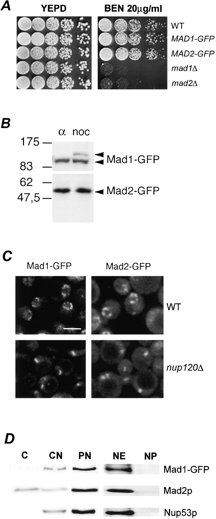
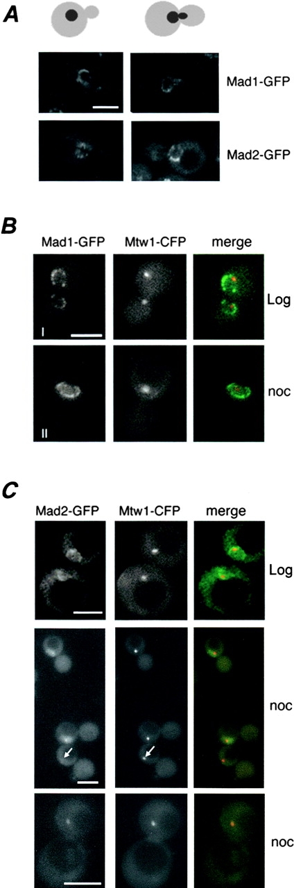
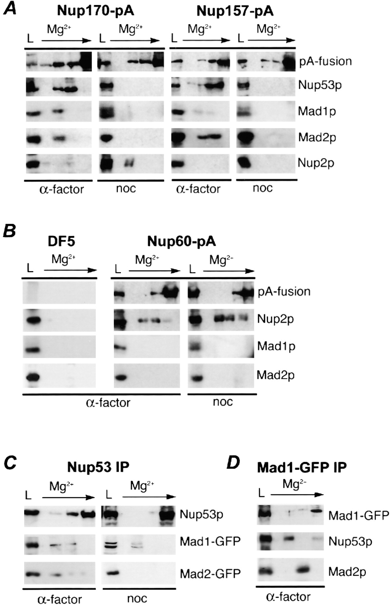
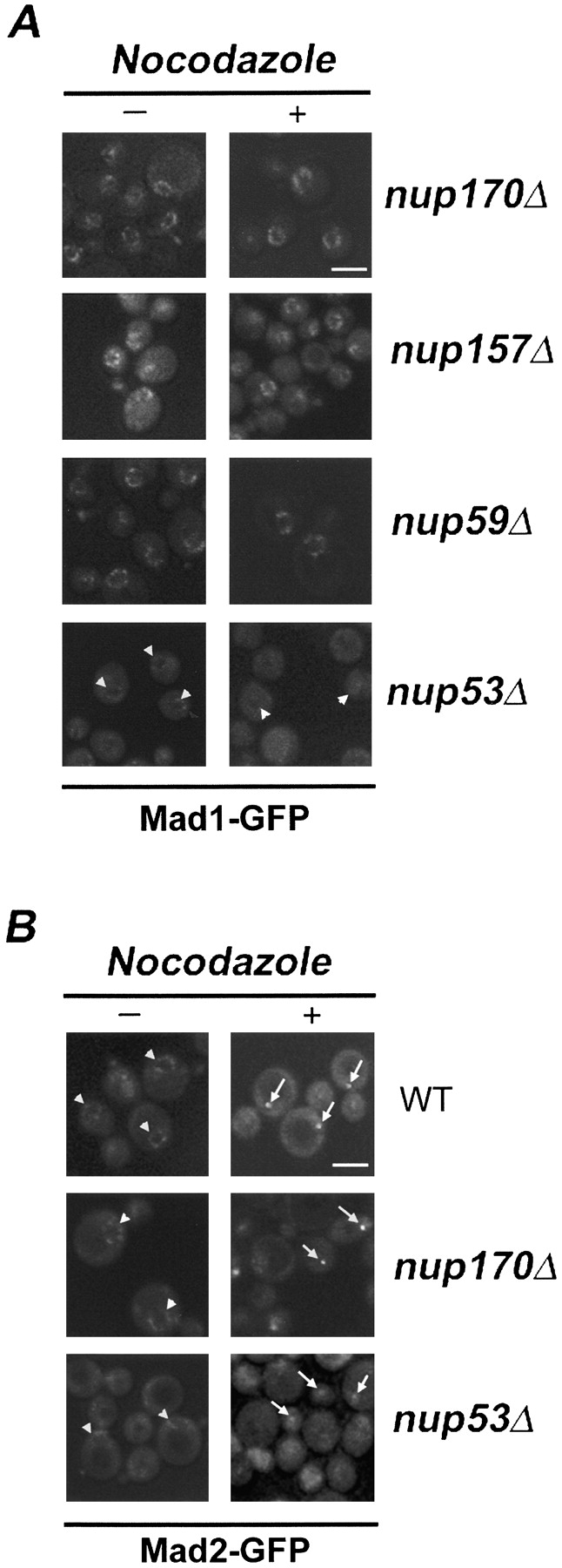
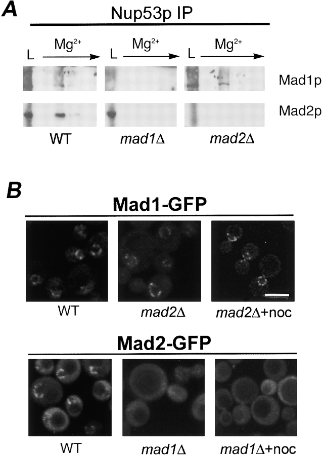
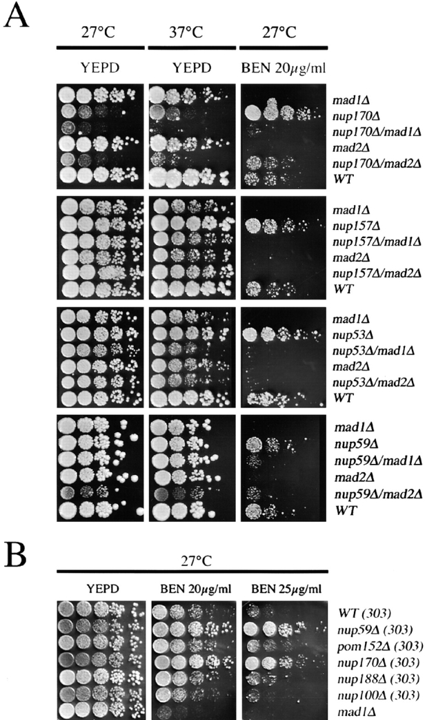
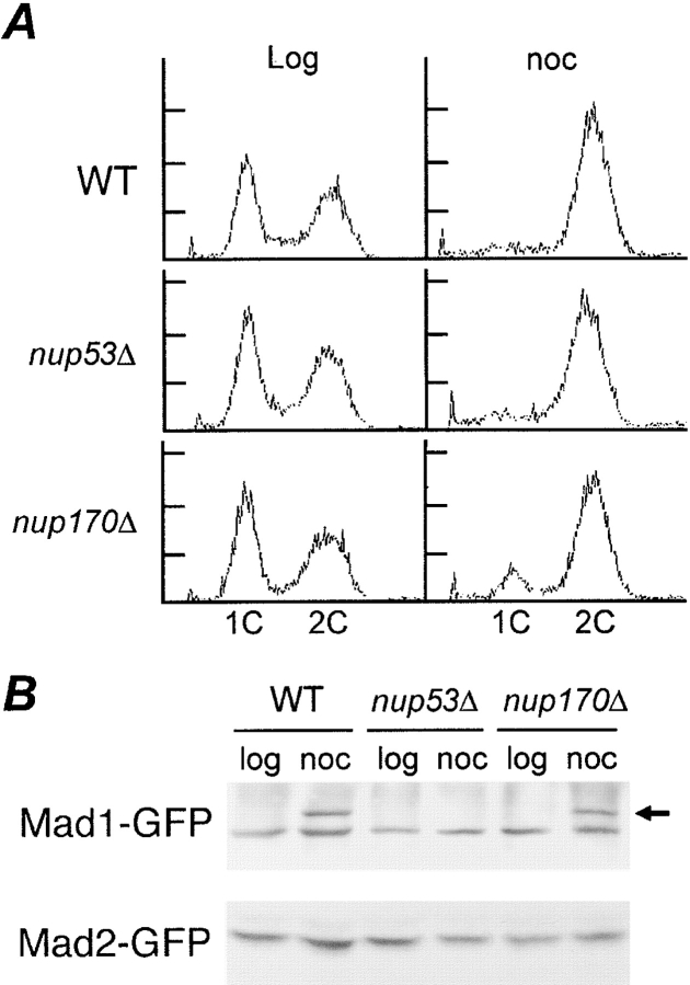
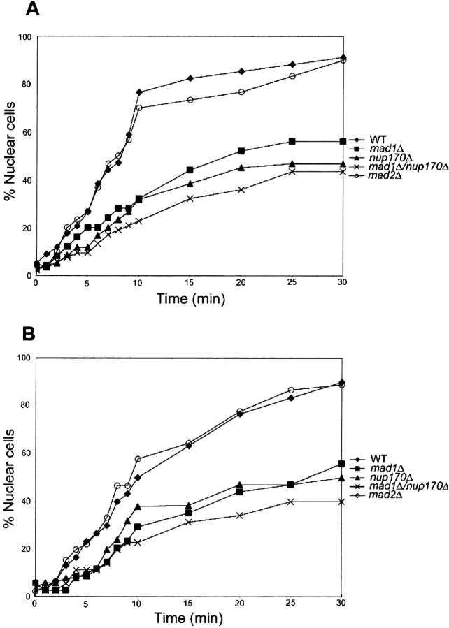
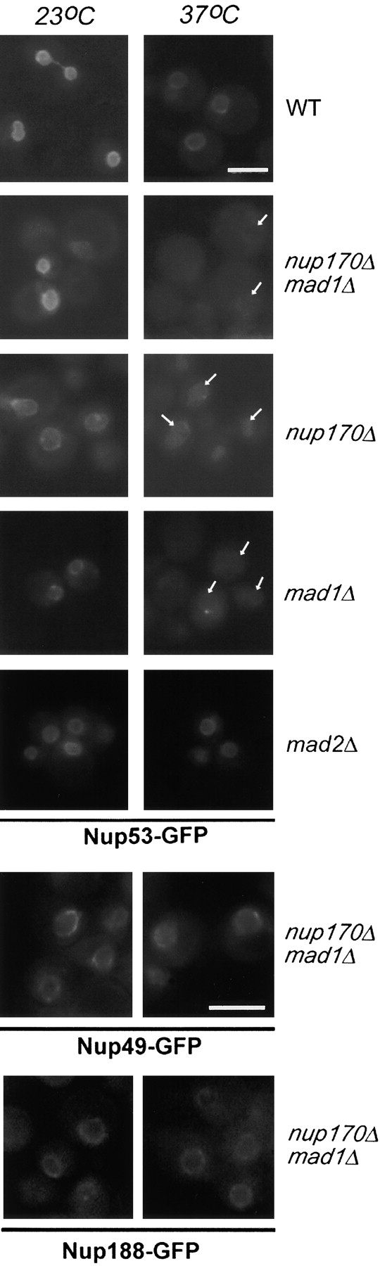
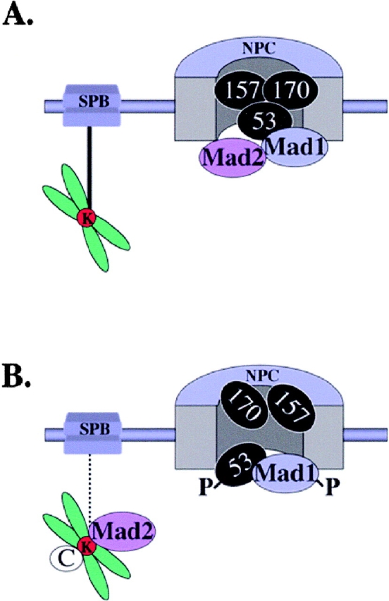
Aphysical and functional link between the nuclear pore complex (NPC) and the spindle checkpoint machinery has been established in the yeast Saccharomyces cerevisiae. We show that two proteins required for the execution of the spindle checkpoint, Mad1p and Mad2p, reside predominantly at the NPC throughout the cell cycle. There they are associated with a subcomplex of nucleoporins containing Nup53p, Nup170p, and Nup157p. The association of the Mad1p-Mad2p complex with the NPC requires Mad1p and is mediated in part by Nup53p. On activation of the spindle checkpoint, we detect changes in the interactions between these proteins, including the release of Mad2p (but not Mad1p) from the NPC and the accumulation of Mad2p at kinetochores. Accompanying these events is the Nup53p-dependent hyperphosphorylation of Mad1p. On the basis of these results and genetic analysis of double mutants, we propose a model in which Mad1p bound to a Nup53p-containing complex sequesters Mad2p at the NPC until its release by activation of the spindle checkpoint. Furthermore, we show that the association of Mad1p with the NPC is not passive and that it plays a role in nuclear transport.


Figures

Figure 1.
Figure 1.
Association of Mad1-GFP and Mad2-GFP with NPCs. (A) Cells expressing Mad1-GFP and Mad2-GFP are not sensitive to the microtubule-destabilizing drug benomyl. The parental strain (WT), MAD1-GFP, MAD2-GFP, mad1Δ, and mad2Δ strains were grown to logarithmic phase in YEPD at 30°C, diluted, and spotted in 10-fold increments on YEPD and YEPD containing 20 μg/ml benomyl (BEN) and incubated for 4 d at 27°C. (B) Western blot analysis of Mad1-GFP– and Mad2-GFP–producing cells. Nuclear extracts were isolated from MAD1-GFP and MAD2-GFP stains arrested in G1 with α-factor (α) or G2/M with nocodazole (noc), and proteins were separated by double-inverted gradient PAGE. Western blots were performed using anti-GFP antibodies. The positions of molecular mass markers are indicated on the left in kD. (C) Mad1-GFP and Mad2-GFP are associated with NPCs. WT or nup120Δ stains synthesizing Mad1-GFP or Mad2-GFP were grown to logarithmic phase in YEPD at 30°C and examined by confocal fluorescence microscopy. Bar, 5 μm. (D) Mad1p and Mad2p are associated with isolated nuclear fractions. Western blot analysis was performed on subcellular fractions derived from the MAD1-GFP strain using anti-GFP, anti-Mad2p, and anti-Nup53p antibodies. Fractions are defined as follows: C, cytosol; CN, crude nuclei; PN, purified nuclei; NE, nuclear envelope pellet; NP, nucleoplasmic fraction. The materials loaded in the C and CN lanes were derived from equal cell equivalents and PN, NE, and NP from 10-fold higher cell equivalents.
Figure 2.
Figure 2.
Spindle checkpoint activation induces the release of Mad2p-GFP, but not Mad1p, from the NPC and its recruitment to kinetochores. (A) Localization of Mad1-GFP and Mad2-GFP to NPCs throughout the cell cycle. Strains producing Mad1-GFP or Mad2-GFP were grown to logarithmic phase in YEPD at 30°C and examined by fluorescence microscopy. Representative images of cells in late S-phase (left) and M-phase (right) are shown. Corresponding shapes of cells (gray) and nuclei (black) are indicated. (B and C) Colocalization of Mad2-GFP and Mtw1-CFP to kinetochores on spindle checkpoint activation. Strains synthesizing the kinetochore marker Mtw1-CFP and either Mad1-GFP (B) or Mad2-GFP (C) were grown to early logarithmic phase of growth (control) or G2/M arrested with nocodazole (noc) and visualized by fluorescence microscopy. Subsequently, images were merged. G2/M arrest of these strains was confirmed by FACS® analysis (not depicted). Two separate magnifications of the Mad2-GFP/Mtw1-CFP cells are shown. In arrested cells, we observed Mtw1-CFP in 75% of the cells and Mad2-GFP foci in 50% of the cells. 94% of cells displaying both signals showed overlapping foci. Note, no bleed through of the Mtw1-CFP signal is observed in the GFP images (arrows). Bars, 5 μm.
Figure 3.
Figure 3.
Mad1p and Mad2p associate with a specific subset of nucleoporins. (A) Nup170-pA or Nup157-pA was affinity-purified using IgG-Sepharose from nuclear extracts isolated from α-factor (α-factor)– and nocodazole (noc)-treated cells. Proteins were eluted with a MgCl2 step gradient and analyzed by Western blotting to detect Mad1p, Mad2p, Nup53p, Nup2p, and the pA fusion. The load fraction (L) is shown. (B) Similar experiments were performed with an untagged WT strain (DF5) and a strain synthesizing Nup60-pA. (C) Mad1p, but not Mad2p, is associated with Nup53p in nocodazole-treated cells. Nup53p was immunoprecipitated using Nup53p- specific antibodies out of nuclear extracts derived from α-factor (α-factor)– and nocodazole (noc)-treated cells synthesizing Mad1-GFP or Mad2-GFP. (D) Nup53p and Mad2p are associated with immunoprecipitated Mad1p. Mad1-GFP was immunoprecipitated from nuclear extracts of α-factor (α-factor)–treated cells synthesizing Mad1-GFP. In C and D, bound complexes were eluted and analyzed as described in A using antibodies direct against GFP (for all GFP fusions), Nup53p, and Mad2p.
Figure 4.
Figure 4.
The effects of nup mutations on the subcellular distribution of the Mad1p–Mad2p complex. (A) The subcellular localization of Mad1-GFP in logarithmically growing (−) or nocodazole-treated (+) nup170Δ, nup157Δ, nup59Δ, and nup53Δ strains was examined using confocal fluorescence microscopy. (B) The localization of Mad2-GFP in logarithmically growing (−) or nocodazole-treated (+) WT, nup170Δ, and nup53Δ strains was examined using confocal fluorescence microscopy. Note, arrowheads point to the NE and arrows point to kinetochore signals. Bars, 5 μm.
Figure 5.
Figure 5.
Mad1p links the Mad1p–Mad2p complex to the NPC. (A) The association of Nup53p with the Mad1p–Mad2p complex requires Mad1p. Nup53p was immunoprecipitated from nuclear extracts of WT, mad1Δ, and mad2Δ strains. Proteins were eluted with a MgCl2 gradient and analyzed by Western blotting with antibodies directed against Mad1p or Mad2p. The load fraction (L) is shown. (B) Mislocalization of Mad2-GFP in a mad1Δ strain. The localization of Mad1-GFP and Mad2-GFP was examined in WT, mad2Δ, or mad1Δ and nocodazole-treated mad2Δ or mad1Δ (+noc) strains using confocal fluorescence microscopy. Bar, 5 μm.
Figure 6.
Figure 6.
MAD1, MAD2, and nup-encoding genes of the Nup53p-containing complex interact genetically. (A) The growth of WT, mad1Δ, mad2Δ, nup170Δ, nup157Δ, nup53Δ, and nup59Δ strains and combinations of double mutants were assessed at 27, 37, and 30°C (not depicted). Strains were grown to early logarithmic phase in YEPD at 30°C, diluted, spotted on YEPD and YEPD containing 20 μg/ml benomyl (BEN), and incubated at the indicated temperatures (27 or 37°C). (B) Benomyl resistance of nup mutants. nup59Δ, pom152Δ, nup170Δ, nup188Δ, nup100Δ, and mad1Δ strains, using W303 as the parental WT strain (WT 303), were spotted on YEPD plates containing either 20 or 25 μg/ml of benomyl and incubated for 3 d at 27°C.
Figure 7.
Figure 7.
Requirement of Nup53p for hyperphosphorylation of Mad1p. (A) nup170Δ and nup53Δ strains arrest in G2/M after treatment with nocodazole. FACS® analysis was performed on logarithmically growing (log) and nocodazole (noc)-treated (1.5 h) WT, nup53Δ, and nup170Δ strains. The positions of 1C and 2C DNA peaks are indicated. (B) Mad1p is not hyperphosphorylated in a nup53Δ strain. Logarithmically growing WT, nup53Δ, and nup170Δ strains expressing either Mad1-GFP or Mad2-GFP were treated with (noc) or without (log) nocodazole for 1.5 h. Total cell lysates were analyzed by immunoblotting using an anti-GFP antibody. The position of hyperphosphorylation Mad1-GFP is indicated by an arrow.
Figure 8.
Figure 8.
Nuclear accumulation of Pho4-GFP is inhibited in strains lacking MAD1 and NUP170. Each of the indicated strains expressing a PHO4-GFP reporter were grown at 30°C (A) or at 30°C and then shifted to 37°C for 3 h (B). After treatment with and removal of metabolic poisons (2-deoxyglucose and sodium azide), relative rates of import were determined by counting the number of cells showing a nuclear accumulation of the reporter and plotting this versus time. The results shown are representative of those obtained in multiple experiments.
Figure 9.
Figure 9.
Mad1p is required for the stable association of Nup53p with the NE. WT, nup170Δmad1Δ, nup170Δ, mad1Δ, and mad2Δ strains expressing a plasmid borne copy of NUP53-GFP (pNP53) were grown to logarithmic phase and either maintained at 23°C or shifted to 37°C for 3 h. The localization of Nup53p-GFP was examined by fluorescence microscopy. The results of similar experiments examining the localization of two other nucleoporins, Nup49-GFP and Nup188-GFP, in the nup170Δ mad1Δ strain are shown in the bottom two rows. Note, arrows point to the NE. Bars, 5 μm.
Figure 10.
Figure 10.
Model summarizing interactions between the Nup53p-containing complex, Mad1p and Mad2p. (A) Shown schematically is an NPC with Nup53p, Nup157p, and Nup170p (53, 157, and 170). The Mad1p–Mad2p complex is associated with the NPC through Mad1p. Additional interactions between Mad proteins and/or the NPC may also exist. Also depicted is one of the spindle pole bodies (SPB) connected to a chromosome's kinetochore (K) via a microtubule (black line). (B) Defects in microtubule interaction (interrupted line) of the SPB with the kinetochore lead to spindle checkpoint activation, hyperphosphorylation of Mad1p (P), dissociation of Mad2p from the NPC, and the recruitment of Mad2p to the kinetochores along with other checkpoint proteins (C). At the same time, Nup157p and Nup170p are no longer associated with phosphorylated Nup53p (P).

References

    1. Adams, A., D.E. Gottschling, C.A. Kaiser, and T. Stearns. 1997. Methods in Yeast Genetics. Cold Spring Harbor Laboratory, Cold Spring Harbor, NY. 177 pp.
    1. Aitchison, J.D., G. Blobel, and M. Rout. 1995. a. Nup120p: a yeast nucleoporin required for NPC distribution and mRNA transport. J. Cell Biol. 131:1659–1675. - PMC - PubMed
    1. Aitchison, J.D., M.P. Rout, M.M. Marelli, G. Blobel, and R.W. Wozniak. 1995. b. Two novel related yeast nucleoporins Nup170p and Nup157p: Complementation with the vertebrate homologue Nup155p and functional interactions with the yeast nuclear pore-membrane protein Pom152p. J. Cell Biol. 131:1133–1148. - PMC - PubMed
    1. Basrai, M.A., J. Kingsbury, D. Koshland, F. Spencer, and P. Hieter. 1996. Faithful chromosome transmission requires Spt4p, a putative regulator of chromatin structure in Saccharomyces cerevisiae Mol. Cell. Biol. 6:2838–2847. - PMC - PubMed
    1. Baudin, A., O. Ozier-Kalogeropoulos, A. Denouel, F. Lacroute, and C. Cullin. 1993. A simple and efficient method for direct gene deletion in Saccharomyces cerevisiae. Nucleic Acids Res. 21:3329–3330. - PMC - PubMed

Publication types

MeSH terms