Comparative Study
. 2002 Dec 9;159(5):833-43.
doi: 10.1083/jcb.200208017. Epub 2002 Dec 9.

The targeting of the atToc159 preprotein receptor to the chloroplast outer membrane is mediated by its GTPase domain and is regulated by GTP

Affiliations
Comparative Study

The targeting of the atToc159 preprotein receptor to the chloroplast outer membrane is mediated by its GTPase domain and is regulated by GTP

Matthew D Smith et al. J Cell Biol. .

Abstract

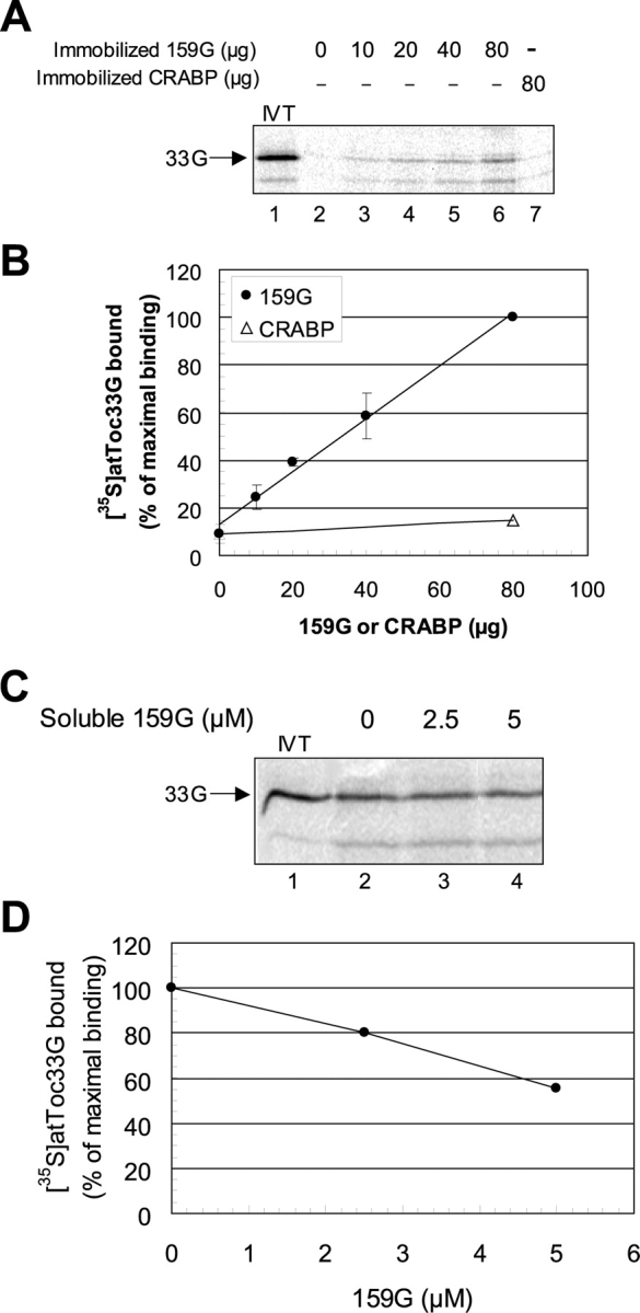
The multimeric translocon at the outer envelope membrane of chloroplasts (Toc) initiates the recognition and import of nuclear-encoded preproteins into chloroplasts. Two Toc GTPases, Toc159 and Toc33/34, mediate preprotein recognition and regulate preprotein translocation. Although these two proteins account for the requirement of GTP hydrolysis for import, the functional significance of GTP binding and hydrolysis by either GTPase has not been defined. A recent study indicates that Toc159 is equally distributed between a soluble cytoplasmic form and a membrane-inserted form, raising the possibility that it might cycle between the cytoplasm and chloroplast as a soluble preprotein receptor. In the present study, we examined the mechanism of targeting and insertion of the Arabidopsis thaliana orthologue of Toc159, atToc159, to chloroplasts. Targeting of atToc159 to the outer envelope membrane is strictly dependent only on guanine nucleotides. Although GTP is not required for initial binding, the productive insertion and assembly of atToc159 into the Toc complex requires its intrinsic GTPase activity. Targeting is mediated by direct binding between the GTPase domain of atToc159 and the homologous GTPase domain of atToc33, the Arabidopsis Toc33/34 orthologue. Our findings demonstrate a role for the coordinate action of the Toc GTPases in assembly of the functional Toc complex at the chloroplast outer envelope membrane.


Figures

Figure 1.
Figure 1.
Energetics of atToc159 targeting to isolated chloroplasts. In vitro–translated [35S]atToc159 was incubated with isolated, energy-depleted chloroplasts in the presence (+) or absence (−) of GTP, ATP, or apyrase for 10 min at 21°C. After the incubation, the reactions were divided equally and one-half was incubated in the presence (+) and the other half in the absence (−) of thermolysin (100 μg/ml) for 30 min on ice. The chloroplasts were reisolated, lysed, and the total membrane fractions analyzed by SDS-PAGE and phosphorimaging. (A) Phosphorimager analysis of SDS-PAGE–resolved chloroplast membranes after the targeting reactions. The results from a typical experiment are shown. Lanes 1 and 7 contain 10% of the [35S]atToc159 in vitro translation product (IVT) added to each reaction. (B) Quantitative analysis of the data from triplicate experiments including those in A. [35S]atToc159 binding to chloroplasts (Binding) was measured as the amount of both full-length atToc159 (atToc159) and 86-kD fragment (159GM) that was associated but not inserted into the outer membrane. The amount of inserted [35S]atToc159 (Insertion) was determined directly from the amount of the 50–55-kD fragments (159M) present after thermolysin treatment. Quantitation of binding and insertion were normalized based on the methionine content of full-length [35S]atToc159 and its fragments. Error bars indicate standard deviation.
Figure 2.
Figure 2.
Effect of nucleotide analogues on the targeting of atToc159 to isolated chloroplasts. The targeting of in vitro translated [35S]atToc159 to isolated chloroplasts in the presence (+) or absence (−) of GTP, GMP-PNP, GDP, GDPβS, or AMP-PNP was performed as described in the legend to Fig. 1. (A) Phosphorimager analysis of SDS-PAGE-resolved chloroplast membranes from the [35S]atToc159 targeting reactions. Lanes 1 and 12 contain 10%, and lanes 6, 9, 17, and 20 contain 20% of the [35S]atToc159 in vitro translation product (IVT) added to each reaction. The positions of full-length atToc159 (atToc159), the 86-kD proteolytic fragment (159GM), and the 50–55 kD thermolysin-resistant fragments (159M) are indicated to the left of the figure. (B) Quantitative analysis of the data from triplicate experiments including those in A. [35S]atToc159 binding (Binding) and insertion (Insertion) were measured as described in the legend to Fig. 1. Error bars indicate standard deviation.
Figure 3.
Figure 3.
Point mutations incorporated into the G-domain of atToc159 and their effects on GTP binding and hydrolysis. (A) Alignment of the GTP binding motifs (G1-G4) of atToc159 with those of human ras (Hs Ras), canine SRP receptor α subunit (Can Srα), E. coli FtsY protein (FtsYp), human SRP54 subunit (Hs SRP54p), and E. coli Ffh protein (Ffhp). The consensus sequences of the G1 through G4 motifs are shown at the top. J, hydrophilic; O, hydrophobic; X, any amino acid. Highly conserved residues are shaded. Arrows indicate the sites of the A864 to R (159-A864R) and K868 to R (159-K868R) point mutations in atToc159. (B) Coomassie-stained SDS-PAGE profile of atToc159G (159G), atToc159G-A864R (159G-A864R), and atToc159G-K868R (159G-K868R) purified by Ni-NTA chromatography from E. coli extracts. Each lane contains 1 μg of protein. The positions of molecular size markers (kD) are indicated to the right of the Figure. (C) GTP binding to wild type and mutant atToc159 G-domains. Purified proteins were bound to nitrocellulose and incubated with 50 nM [α-32P]GTP (3,000 Ci/mmol) in the presence of 1 μM ATP. Bound [α-32P]GTP was quantitated using a phosphorimager. (D) GTP hydrolysis by wild-type and mutant atToc159 G-domains. 1 μM [α-32P]GTP (150 mCi/μmol) was incubated with 0.5 μM atToc159G, atToc159G-A864R or atToc159G-K868R in a 25 μl reaction for 20 min at 25°C. Radiolabeled GTP and GDP were resolved by thin-layer chromatography and radioactivity was quantitated using a phosphorimager. Error bars indicate standard deviation. N.D., not detectable above background.
Figure 3.
Figure 3.
Point mutations incorporated into the G-domain of atToc159 and their effects on GTP binding and hydrolysis. (A) Alignment of the GTP binding motifs (G1-G4) of atToc159 with those of human ras (Hs Ras), canine SRP receptor α subunit (Can Srα), E. coli FtsY protein (FtsYp), human SRP54 subunit (Hs SRP54p), and E. coli Ffh protein (Ffhp). The consensus sequences of the G1 through G4 motifs are shown at the top. J, hydrophilic; O, hydrophobic; X, any amino acid. Highly conserved residues are shaded. Arrows indicate the sites of the A864 to R (159-A864R) and K868 to R (159-K868R) point mutations in atToc159. (B) Coomassie-stained SDS-PAGE profile of atToc159G (159G), atToc159G-A864R (159G-A864R), and atToc159G-K868R (159G-K868R) purified by Ni-NTA chromatography from E. coli extracts. Each lane contains 1 μg of protein. The positions of molecular size markers (kD) are indicated to the right of the Figure. (C) GTP binding to wild type and mutant atToc159 G-domains. Purified proteins were bound to nitrocellulose and incubated with 50 nM [α-32P]GTP (3,000 Ci/mmol) in the presence of 1 μM ATP. Bound [α-32P]GTP was quantitated using a phosphorimager. (D) GTP hydrolysis by wild-type and mutant atToc159 G-domains. 1 μM [α-32P]GTP (150 mCi/μmol) was incubated with 0.5 μM atToc159G, atToc159G-A864R or atToc159G-K868R in a 25 μl reaction for 20 min at 25°C. Radiolabeled GTP and GDP were resolved by thin-layer chromatography and radioactivity was quantitated using a phosphorimager. Error bars indicate standard deviation. N.D., not detectable above background.
Figure 4.
Figure 4.
The effects of GTPase mutations on the targeting of atToc159 to isolated chloroplasts. The targeting of in vitro–translated [35S]atToc159 (atToc159), [35S]atToc159-A864R (159-A864R), or [35S]atToc159-K868R (159-K868R) to isolated chloroplasts in the presence of GTP or GDP was performed as described in the legend of Fig. 1. (A) Phosphorimager analysis of SDS-PAGE-resolved chloroplast membranes from targeting reactions performed in the presence of GTP. Lanes 1, 4, and 7 contain 10% of the [35S]atToc159, [35S]atToc159-A864R, and [35S]atToc159-K868R in vitro translation products (IVT) added to each reaction, respectively. (B) Quantitative analysis of the data from triplicate experiments including those in A. [35S]atToc159 binding (Binding) and insertion (Insertion) were measured as described in the legend to Fig. 1. (C) GDP binding to wild-type and mutant atToc159-A864R G-domains. Binding of [3H]GDP (32 Ci/mmol) to purified atToc159G and atToc159G-A864R was measured in a filter binding assay in the presence of ATP. Bound [3H]GDP was quantitated by scintillation counting. (D) Phosphorimager analysis of SDS-PAGE–resolved chloroplast membranes from time courses of atToc159 and atToc159-A864R insertion performed in the presence of GTP or GDP. Lanes 1 and 7 contain 20% of the [35S]atToc159 (top) and [35S]atToc159-A864R (bottom) in vitro translation products (IVT) added to each reaction. The results from a typical experiment are shown. (E) Quantitative analysis of the data from duplicate experiments including those in D. Insertion of [35S]atToc159 was measured as described in the legend to Fig. 1. The positions of the atToc159 proteins, the 86-kD proteolytic fragments (159GM) and the 50–55-kD thermolysin- protected fragments (159M) are indicated to the left of the figures. Error bars indicate standard deviation.
Figure 4.
Figure 4.
The effects of GTPase mutations on the targeting of atToc159 to isolated chloroplasts. The targeting of in vitro–translated [35S]atToc159 (atToc159), [35S]atToc159-A864R (159-A864R), or [35S]atToc159-K868R (159-K868R) to isolated chloroplasts in the presence of GTP or GDP was performed as described in the legend of Fig. 1. (A) Phosphorimager analysis of SDS-PAGE-resolved chloroplast membranes from targeting reactions performed in the presence of GTP. Lanes 1, 4, and 7 contain 10% of the [35S]atToc159, [35S]atToc159-A864R, and [35S]atToc159-K868R in vitro translation products (IVT) added to each reaction, respectively. (B) Quantitative analysis of the data from triplicate experiments including those in A. [35S]atToc159 binding (Binding) and insertion (Insertion) were measured as described in the legend to Fig. 1. (C) GDP binding to wild-type and mutant atToc159-A864R G-domains. Binding of [3H]GDP (32 Ci/mmol) to purified atToc159G and atToc159G-A864R was measured in a filter binding assay in the presence of ATP. Bound [3H]GDP was quantitated by scintillation counting. (D) Phosphorimager analysis of SDS-PAGE–resolved chloroplast membranes from time courses of atToc159 and atToc159-A864R insertion performed in the presence of GTP or GDP. Lanes 1 and 7 contain 20% of the [35S]atToc159 (top) and [35S]atToc159-A864R (bottom) in vitro translation products (IVT) added to each reaction. The results from a typical experiment are shown. (E) Quantitative analysis of the data from duplicate experiments including those in D. Insertion of [35S]atToc159 was measured as described in the legend to Fig. 1. The positions of the atToc159 proteins, the 86-kD proteolytic fragments (159GM) and the 50–55-kD thermolysin- protected fragments (159M) are indicated to the left of the figures. Error bars indicate standard deviation.
Figure 5.
Figure 5.
The effects of deletion mutations on the targeting of atToc159 to isolated chloroplasts. The targeting of in vitro translated [35S]atToc159 (atToc159), [35S]atToc159GM (159GM), [35S]atToc159A (159A), [35S]atToc159M (159M), or [35S]atToc159G (159G) to isolated chloroplasts in the presence of ATP and GTP was performed as described in the legend to Fig. 1. (A) Phosphorimager analysis of SDS-PAGE-resolved chloroplast membranes from the targeting reactions. Lanes 1, 4, 7, 10, and 13 contain 10% of the [35S]atToc159, [35S]atToc159GM, [35S]atToc159A, [35S]atToc159M, and [35S]atToc159G in vitro translation products (IVT) added to each reaction, respectively. The positions of atToc159, 159A, 159GM, 159M, and 159G are indicated to the left of the figure. Results from a typical experiments are shown. (B) Quantitative analysis of the data from triplicate experiments including those in A. Binding and insertion of the [35S]atToc159 deletion constructs were measured as described in the legend to Fig. 1. Error bars indicate standard deviation.
Figure 6.
Figure 6.
Binding of atToc159G to isolated chloroplasts. (A) In vitro–translated [35S]atToc159G (159G) was incubated with isolated chloroplasts in a standard targeting assay (Fig. 1, legend) in either the absence or presence of increasing concentrations of purified, unlabeled atToc159G. The total membrane fraction from each targeting reaction was resolved by SDS-PAGE and analyzed using a phosphorimager. 20% of the [35S]atToc159G in vitro translation product (IVT) added to each reaction is shown in lane 1. (B) Quantitative analysis of the data from triplicate experiments including those in A. (C) Competition of purified atToc159G (159G) with atToc159 for targeting to chloroplasts. [35S]atToc159 was incubated with isolated, intact chloroplasts in a standard targeting assay (Fig. 1, legend) in the absence (−) or presence (+) of 0.25 μM purified atToc159G. Total membrane fractions were separated by SDS-PAGE and analyzed using a phosphorimager as described in the legend to Fig. 1. Lanes 1 and 4 contain 10% of the [35S]atToc159 in vitro translation product (IVT) added to each reaction. The positions of full-length atToc159 (atToc159), the 86-kD proteolytic fragment (159GM), and the 50–55-kD thermolysin-resistant fragments (159M) are indicated to the left of the figure. (D) Quantitative analysis of the data from triplicate experiments including those in C. [35S]atToc159 binding (Bound) and insertion (Inserted) were measured as described in the legend to Fig. 1. Error bars indicate standard deviation.
Figure 7.
Figure 7.
Binding of wild-type and mutant atToc159 G-domains to chloroplasts in vitro. (A) In vitro–translated [35S]atToc159G (159G), [35S]atToc159G-A864R (159G-A864R), or [35S]atToc159G-K868R (159G-K868R) was incubated with isolated chloroplasts in a standard targeting assay (Fig. 1, legend). The total membrane fraction from each targeting reaction (lanes 2, 4, and 6) was resolved by SDS-PAGE and analyzed using a phosphorimager. 10% of the [35S]atToc159G, [35S]atToc159G-A864R, or [35S]atToc159G-K868R in vitro translation product (IVT) added to each reaction is shown in lanes 1, 3, and 5, respectively. (B) Quantitative analysis of the data from triplicate experiments including those in A. (C) Competition of purified atToc159G-A864R and atToc159G-K868R with atToc159 for targeting to chloroplasts. [35S]atToc159 was incubated with isolated, intact chloroplasts in a standard targeting assay (Fig. 1, legend) in the absence (−) or presence (+) of 0.25 μM purified 159G-A864R or 159G-K868R. Total membrane fractions were separated by SDS-PAGE and analyzed using a phosphorimager as described in the legend to Fig. 1. Lane 1 contains 10% of the [35S]atToc159 in vitro translation product (IVT) added to each reaction. (D) Quantitative analysis of the data from triplicate experiments including those in C. Error bars indicate standard deviation.
Figure 8.
Figure 8.
Direct binding of atToc33G to atToc159G. (A) [35S]atToc33G was incubated in the presence of GTP with the indicated amounts of hexahistidine-tagged atToc159G (159G) or cellular retinoic acid binding protein (CRABP) that had been immobilized on Ni-NTA resin. Bound proteins were eluted and separated by SDS-PAGE and analyzed using a phosphorimager. Lane 1 contains 20% of the [35S]atToc33G added to each reaction. (B) Quantitation of the data presented in A. Error bars represent standard deviation. (C) Competition of soluble atToc159G with immobilized atToc159G for binding to atToc33G. [35S]atToc33G was incubated with immobilized hexahistidine-tagged atToc159G in the absence or presence of increasing concentrations of soluble atToc159G. (D) Quantitation of the data presented in C.
Figure 9.
Figure 9.
Energetics of atToc33G-atToc159G binding. Nucleotide-depleted in vitro–translated [35S]atToc33G was incubated with immobilized hexahistidine-tagged atToc159G (159G) in the absence (−) or presence (+) of 0.1 mM GTP, GMP-PNP, ATP, AMP-PNP, GDP, or GDPβS. The bound proteins were eluted and separated by SDS-PAGE and analyzed using a phosphorimager. (A) Phosphorimager analysis of bound [35S]atToc33G. 10% of the in vitro–translated [35S]atToc33G (IVT) that was added to each reaction is shown in lane 1. Lane 2 contains the [35S]atToc33G that bound to the Ni-NTA matrix in the absence of atToc159G. (B) Quantitation of the data presented in A.

Similar articles

Cited by

References

    1. Akita, M., E. Nielsen, and K. Keegstra. 1997. Identification of protein transport complexes in the chloroplastic envelope membranes via chemical cross-linking. J. Cell Biol. 136:983–994. - PMC - PubMed
    1. Althoff, S.M., S.W. Stevens, and J.A. Wise. 1994. The Srp54 GTPase is essential for protein export in the fission yeast Schizosaccharomyces pombe. Mol. Cell. Biol. 14:7839–7854. - PMC - PubMed
    1. Arnon, D.I. 1949. Copper enzymes in isolated chloroplasts. Polyphenooxidase in Beta vulgaris. Plant Physiol. 24:1–15. - PMC - PubMed
    1. Bauer, J., K. Chen, A. Hiltbunner, E. Wehrli, M. Eugster, D. Schnell, and F. Kessler. 2000. The major protein import receptor of plastids is essential for chloroplast biogenesis. Nature. 403:203–207. - PubMed
    1. Bauer, J., A. Hiltbrunner, and F. Kessler. 2001. Molecular biology of chloroplast biogenesis: gene expression, protein import and intraorganellar sorting. Cell. Mol. Life Sci. 58:420–433. - PMC - PubMed

Publication types

MeSH terms