Comparative Study
. 2002 Dec 9;159(5):867-80.
doi: 10.1083/jcb.200209039. Epub 2002 Dec 9.

Lineage-specific requirements of beta-catenin in neural crest development

Affiliations
Comparative Study

Lineage-specific requirements of beta-catenin in neural crest development

Lisette Hari et al. J Cell Biol. .

Abstract

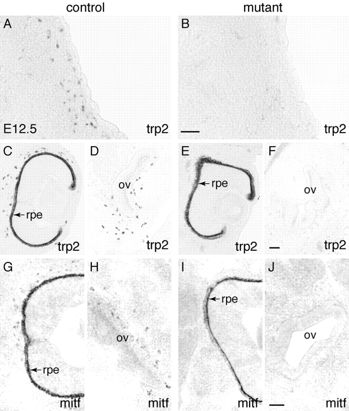
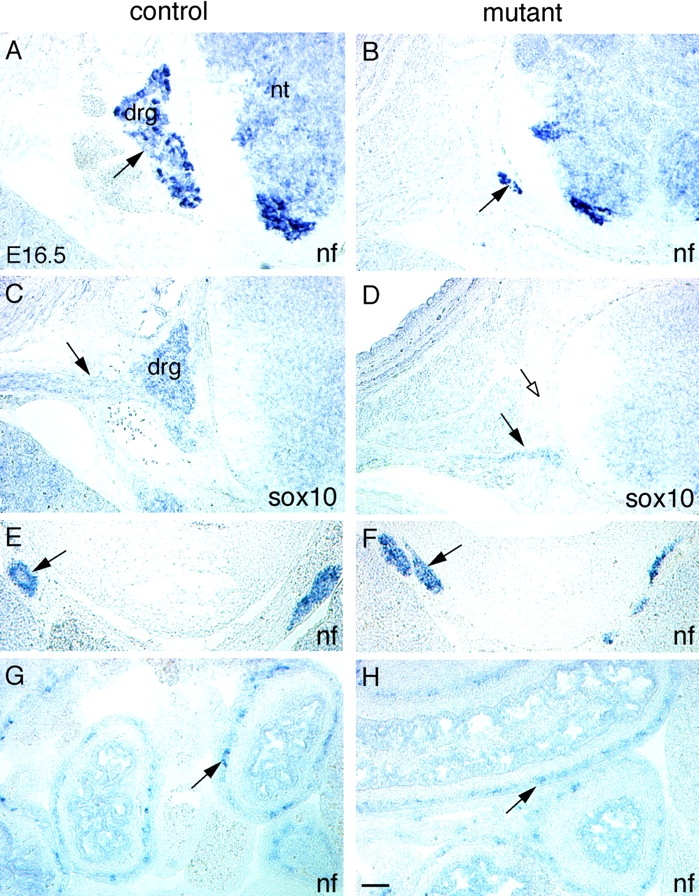
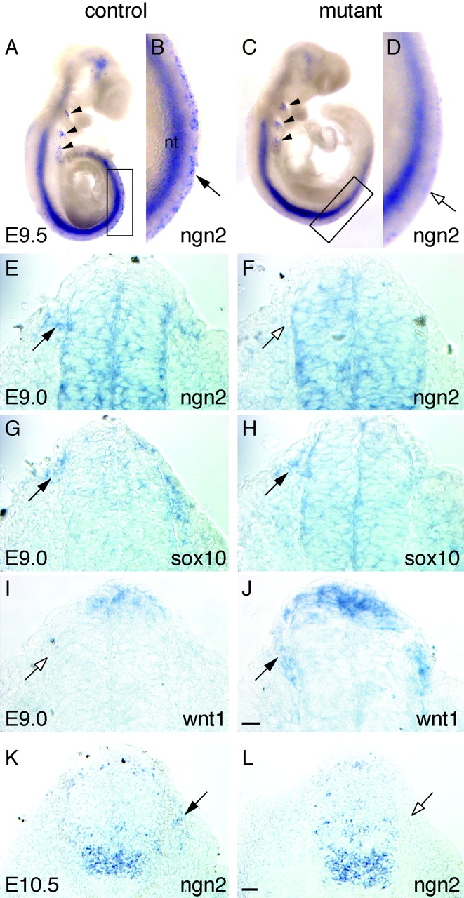
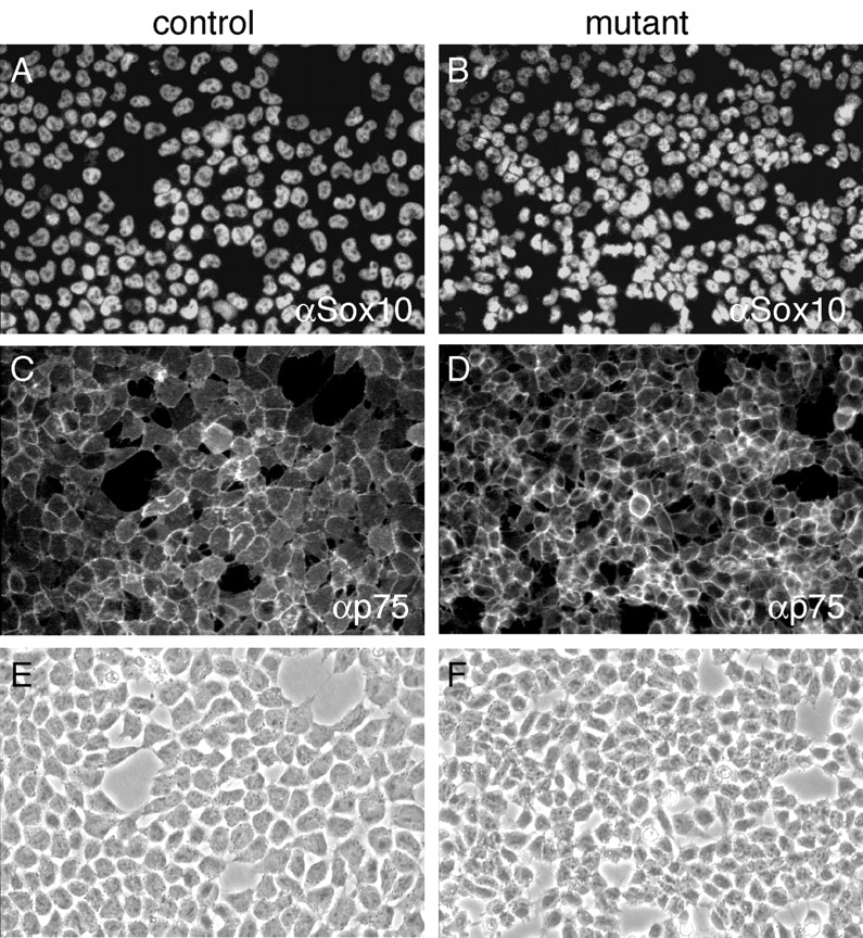
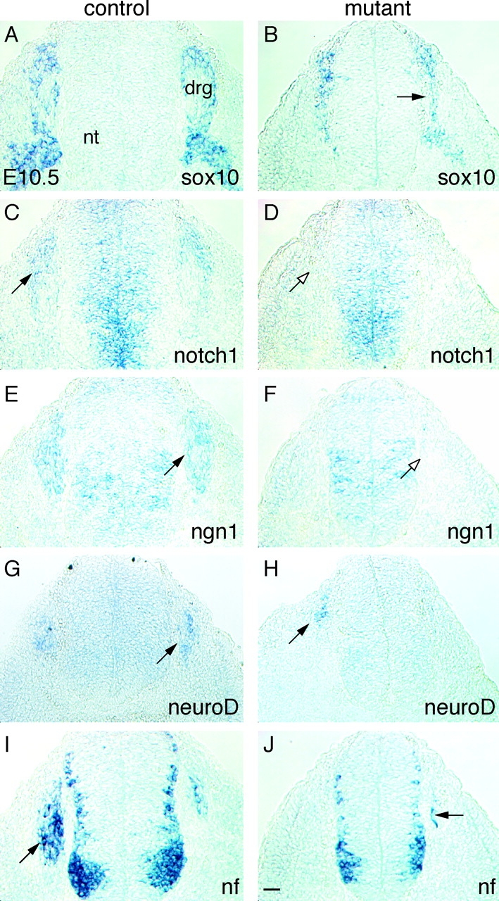
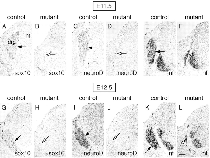
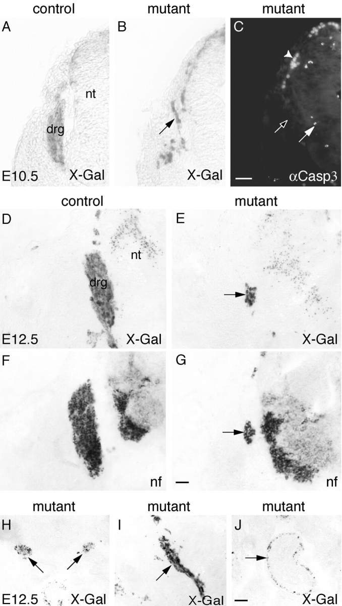
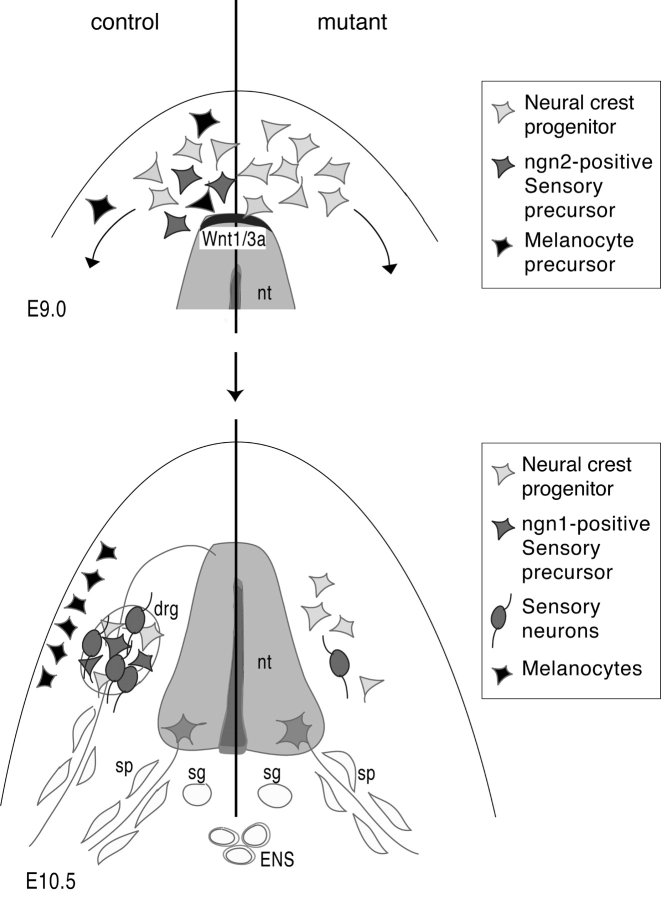
Beta-catenin plays a pivotal role in cadherin-mediated cell adhesion. Moreover, it is a downstream signaling component of Wnt that controls multiple developmental processes such as cell proliferation, apoptosis, and fate decisions. To study the role of beta-catenin in neural crest development, we used the Cre/loxP system to ablate beta-catenin specifically in neural crest stem cells. Although several neural crest-derived structures develop normally, mutant animals lack melanocytes and dorsal root ganglia (DRG). In vivo and in vitro analyses revealed that mutant neural crest cells emigrate but fail to generate an early wave of sensory neurogenesis that is normally marked by the transcription factor neurogenin (ngn) 2. This indicates a role of beta-catenin in premigratory or early migratory neural crest and points to heterogeneity of neural crest cells at the earliest stages of crest development. In addition, migratory neural crest cells lateral to the neural tube do not aggregate to form DRG and are unable to produce a later wave of sensory neurogenesis usually marked by the transcription factor ngn1. We propose that the requirement of beta-catenin for the specification of melanocytes and sensory neuronal lineages reflects roles of beta-catenin both in Wnt signaling and in mediating cell-cell interactions.


Figures

Figure 1.
Figure 1.
Absence of melanocytes in β -catenin mutant embryos. Melanocytes and their precursors were marked by in situ hybridization analysis on transverse sections of E12.5 control (A, C, D, G, and H) and mutant (B, E, F, I, and J) embryos using trp2 (A–F) and mitf (G–J) riboprobes. In mutant embryos, trp2-positive melanocytes were absent underneath the surface ectoderm (B), in areas surrounding the retinal pigment epithelium (rpe, E), and around the otic vesicle (ov, F), whereas many melanocytes were present in control embryos (A, C, and D). Many mitf-expressing melanoblasts were found around the eye (G) and the otic vesicle (H) of control but not mutant embryos (I and J). Bars, 50 μm.
Figure 2.
Figure 2.
Analysis of control and mutant PNS. In situ hybridization experiments on transverse sections of E16.5 embryos with nf (A and B and E–H) and sox10 (C and D) riboprobes revealed a reduction of neuronal (A and B, arrows) and complete absence of glial (C and D, open arrow) lineages in dorsal root ganglia (drg). Peripheral nerves marked by sox10 (C and D, arrows) were reduced in diameter, whereas other crest derivatives, such as sympathetic ganglia (E and F, arrows) and the enteric nervous system (G and H, arrows), appeared to develop normally. nt, neural tube. Bar, 50 μm.
Figure 3.
Figure 3.
Absence of the ngn2-expressing sensory sublineage in the emigrating crest of mutant embryos. Whole mount in situ hybridization experiments showed ngn2 expression in the neural tube and in placodes (arrowheads) of control and mutant embryos at E9.5 (A–D). In migratory crest, ngn2 mRNA was only detectable in control (A and B, arrow) but not in mutant embryos (C and D, open arrow). The boxes in A and C indicate the areas enlarged in B and D, respectively. On transverse sections at E9.0, ngn2-positive neural crest cells were found in control (E, arrow) but not in mutant (F, open arrow) embryos. In contrast, on adjacent sections, neural crest emigrating from the dorsal neural tube was marked by sox10 mRNA both in control and mutant embryos (G and H, arrows). As at earlier stages, ngn2 mRNA was present in the DRG anlage of control (K, arrow) but not of mutant embryos (L, open arrow) at E10.5. Hybridization with a wnt1 riboprobe showed maintained wnt1 expression in the emerging crest of mutant embryos (J, arrow), whereas it was down-regulated in neural crest of control embryos (I, open arrow) at E9.0. Bars: (E–J) 10 μm; (K and L) 20 μm.
Figure 4.
Figure 4.
Normal emigration of mutant neural crest cells. Neural crest explants were obtained from neural tubes that had been isolated from control and β-catenin mutant mice at E9 and cultured for 20 h to allow emigration of neural crest cells. After emigration, neural crest cells were fixed and immunolabeled with anti-sox10 antibody (visualized by Cy3 fluorescence) (A and B) and anti-p75 antibody (visualized by FITC fluorescence) (C and D). Note that virtually all neural crest cells were double positive for the neural crest stem cell markers p75 and sox10. (E and F) Corresponding phase contrast pictures. (G) To compare and quantify the extent of control and mutant neural crest outgrowth after 20 h, the migration index was calculated using the NIH image 1.62 software (Materials and methods). Two independent experiments using nonsibling embryos were performed, scoring three explants of control and mutant embryos per experiment. Each bar represents the migration index (mean ± SD) of three different explants. Note that mutant explants were not significantly reduced in size and density.
Figure 4.
Figure 4.
Normal emigration of mutant neural crest cells. Neural crest explants were obtained from neural tubes that had been isolated from control and β-catenin mutant mice at E9 and cultured for 20 h to allow emigration of neural crest cells. After emigration, neural crest cells were fixed and immunolabeled with anti-sox10 antibody (visualized by Cy3 fluorescence) (A and B) and anti-p75 antibody (visualized by FITC fluorescence) (C and D). Note that virtually all neural crest cells were double positive for the neural crest stem cell markers p75 and sox10. (E and F) Corresponding phase contrast pictures. (G) To compare and quantify the extent of control and mutant neural crest outgrowth after 20 h, the migration index was calculated using the NIH image 1.62 software (Materials and methods). Two independent experiments using nonsibling embryos were performed, scoring three explants of control and mutant embryos per experiment. Each bar represents the migration index (mean ± SD) of three different explants. Note that mutant explants were not significantly reduced in size and density.
Figure 5.
Figure 5.
Mutant neural crest cells are unable to generate sensory neurons. Neural crest explants from control and mutant mice were prepared as described in the legend to Fig. 4. The cells were allowed to differentiate and were fixed after 36 h (A–D) or 48 h (E–H) in culture. The cultures were immunolabeled using anti–Brn-3A antibody (visualized by Cy3 fluorescence) (A, B, E, and F) and double stained either with anti-sox10 antibody (visualized by FITC fluorescence) (A and B) or anti–nf 160 (NF) (visualized by FITC fluorescence) (E and F). Sensory neuron precursors, defined by the expression of Brn-3A (A, arrow), and sensory neurons, defined by coexpression of Brn-3A and NF (E, arrow), were completely absent in mutant explants (B and F). Note that a few Brn-3A–negative nonsensory neurons were found in mutant explants (F, arrowhead). (C, D, G, and H) Corresponding phase contrast pictures.
Figure 6.
Figure 6.
Failure of DRG formation from sox10-expressing neural crest–derived progenitor cells. Sox10-expressing cells were present in condensing DRG of control embryos (A) and lateral to the neural tube (nt) of mutant embryos (B, arrow) at E10.5. Near-adjacent sections hybridized with Notch1 riboprobes demonstrate lack of notch1 expression in sox10-positive progenitors of mutant embryos (D, open arrow), whereas Notch1 is expressed in progenitors of control embryos (C, arrow). Furthermore, ngn1-expressing cells were virtually absent lateral to the neural tube of mutant embryos (F, open arrow), whereas extensive ngn1 expression was found in the forming DRG of control embryos (E, arrow). In contrast to control embryos (G and I, arrows), only a few neuroD- and nf-expressing cells were present in mutant embryos (H and J, arrows). Bar, 20 μm.
Figure 7.
Figure 7.
Absence of de novo neurogenesis in the DRG anlage of mutant embryos at later stages. At E11.5 (A–F) and E12.5 (G-L), progenitors and glial cells detected by hybridization with a sox10 riboprobe (A and G, arrows), as well as neurons and their precursors detected by expression of nf (E and K, arrows) and neuroD (C and I, arrows) mRNA, respectively, constituted the DRG in control embryos. In contrast, sox10- (B and H, open arrows) and neuroD- (D and J, open arrows) positive cells were completely missing in the mutant. Moreover, only a few nf-expressing neurons were present in mutant embryos (F and L, open arrows). Bar, 50 μm.
Figure 8.
Figure 8.
Survival and in vivo fate mapping of mutant cells. Control embryos carrying wnt1-Cre and the R26R allele displayed β-galactosidase activity in the developing DRG at E10.5 (A) and E12.5 (D). At E12.5, the control DRG was outlined by nf expression (F). Cre-expressing neural crest cells and their derivatives were also detectable by β-galactosidase expression in mutant embryos carrying wnt1-Cre and the R26R allele. At E10.5, mutant cells were localized lateral to the neural tube without forming proper DRG (B). No increased cell death was found in this area, as shown by immunostaining for the activated form of caspase3 (Casp3) (C, open arrow). B and C are adjacent sections. Note the Casp3-positive cells within the neural tube (arrow) and autofluorescent blood cells present at the dorsal margins of the embryo (arrowhead). At E12.5, neural crest–derived mutant cells expressing β-galactosidase (E, arrow) were confined to the domain of a few nf-positive cells present in the mutant (G, arrow). E and G represent adjacent sections. Mutant cells were able to normally colonize other neural crest–derived structures, such as sympathetic ganglia (H, arrows), peripheral nerves (I, arrow), and the enteric nervous system (J, arrow). Bars: (A–C) 50 μm; (D–J) 100 μm.
Figure 9.
Figure 9.
Lineage-specific requirement of β -catenin in neural crest development. (E9.0, control) Emigrating neural crest appears to be heterogeneous, consisting of multipotent neural crest progenitor cells, early sensory precursors (marked by ngn2 expression), and possibly melanoblasts. (E9.0, β-catenin mutant) Neural crest fails to generate sensory precursors and melanoblasts. The specification of sensory and melanocytic fates in premigratory or early migratory neural crest might depend on signaling by wnt1/wnt3a, which are expressed in the dorsal neural tube (nt) at the stage of crest emigration. (E10.5, control) Neural crest–derived progenitor cells aggregate in DRG and produce ngn1-dependent sensory precursors. (E10.5, mutant) Progenitors lateral to the neural tube fail to aggregate and to form proper DRG; virtually no ngn1-expressing sensory precursors and only very few differentiated sensory neurons are detectable. This might point to a role of β-catenin in mediating cell–cell interactions possibly involved in sensory neurogenesis, although other β-catenin functions cannot be excluded. Other crest derivatives such as sympathetic ganglia (sg), the enteric nervous system (ENS), and Schwann cell precursors along peripheral nerves (sp) form independently of β-catenin activity.

References

    1. Anderson, D.J. 1999. Lineages and transcription factors in the specification of vertebrate primary sensory neurons. Curr. Opin. Neurobiol. 9:517–524. - PubMed
    1. Anderson, D.J., A. Groves, L. Lo, Q. Ma, M. Rao, N.M. Shah, and L. Sommer. 1997. Cell lineage determination and the control of neuronal identity in the neural crest. Cold Spring Harb. Symp. Quant. Biol. 62:493–504. - PubMed
    1. Borchers, A., R. David, and D. Wedlich. 2001. Xenopus cadherin-11 restrains cranial neural crest migration and influences neural crest specification. Development. 128:3049–3060. - PubMed
    1. Brault, V., R. Moore, S. Kutsch, M. Ishibashi, D.H. Rowitch, A.P. McMahon, L. Sommer, O. Boussida, and R. Kemler. 2001. Inactivation of the β-catenin gene by Wnt1-Cre-mediated deletion results in dramatic brain malformation and failure of craniofacial development. Development. 128:1253–1264. - PubMed
    1. Cadigan, K.M., and R. Nusse. 1997. Wnt signaling: a common theme in animal development. Genes Dev. 11:3286–3305. - PubMed

Publication types

MeSH terms