

Increased expression of AP2 and Sp1 transcription factors in human thyroid tumors: a role in NIS expression regulation?
- PMID: 12475396
- PMCID: PMC139985
- DOI: 10.1186/1471-2407-2-35
Increased expression of AP2 and Sp1 transcription factors in human thyroid tumors: a role in NIS expression regulation?
Abstract
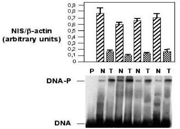
Background: Sodium/iodide symporter (NIS) is a key protein in iodide transport by thyroid cells and this activity is a prerequisite for effective radioiodide treatment of thyroid cancer. In the majority of thyroid cancers, however, iodide uptake is reduced, probably as a result of decreased NIS protein expression.
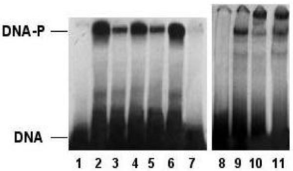
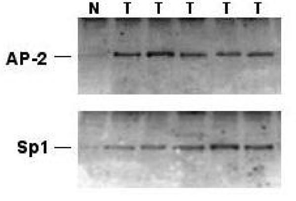
Methods: To identify the mechanisms that negatively affect NIS expression in thyroid tumors, we performed electrophoresis mobility shift assays and immunoblot analysis of nuclear protein extracts from normal and tumoral thyroid tissues from 14 unrelated patients.
Results: Two proteins closely related to the transcription factors AP2 and Sp1 were identified in the nuclear extracts. Expression of both AP2 and Sp1 in nuclear extracts from thyroid tumors was significantly higher than that observed in corresponding normal tissues.
Conclusion: These observations raise the possibility that NIS expression, and subsequently iodide transport, are reduced in thyroid tumors at least in part owing to alterations in the binding activity of AP2 and Sp1 transcription factors to NIS promoter.
Figures
References
-
- Filetti S, Bidart JM, Arturi F, Caillou B, Russo D, Schlumberger M. Sodium/iodide symporter: a key transport system in thyroid cancer cell metabolism. Eur J Endocrinol. 1999;141:443–457. - PubMed
-
- De La Vieja A, Dohan O, Levy O, Carrasco N. Molecular analysis of the sodium/symporter: impact on thyroid and extrathyroid pathophysiology. Physiol Rev. 2000;80:1083–1105. - PubMed
-
- Arturi F, Russo D, Schlumberger M, DuVillard JA, Caillou B, Vigneri P, Wicker R, Chiefari E, Suarez HG, Filetti S. Iodide symporter gene expression in human thyroid tumors. J Clin Endocrinol Metab. 1998;83:2493–2496. - PubMed
-
- Lazar V, Bidart JM, Caillou B, Mahé C, Lacroix L, Filetti S, Schlumberger M. Expression of the Na+/I- symporter gene in human thyroid tumors: a comparison study with other thyroid-specific genes. J Clin Endocrinol Metab. 1999;84:3228–3234. - PubMed
-
- Ryu KY, Tong Q, Jhiang SM. Promoter characterization of the human Na+/I- symporter. J Clin Endocrinol Metab. 1998;83:3247–3251. - PubMed
Publication types
MeSH terms
Substances
Grants and funding
LinkOut - more resources
Full Text Sources
Medical
Molecular Biology Databases
Research Materials
