Review
. 2002 Dec;3(12):1139-45.
doi: 10.1093/embo-reports/kvf246.

Telomere architecture

Affiliations
Review

Telomere architecture

Daniela Rhodes et al. EMBO Rep. 2002 Dec.

Abstract

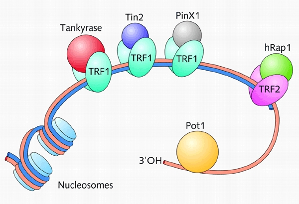
Telomeres are protein-DNA complexes that cap chromosome ends and protect them from being recognized and processed as DNA breaks. Loss of capping function results in genetic instability and loss of cellular viability. The emerging view is that maintenance of an appropriate telomere structure is essential for function. Structural information on telomeric proteins that bind to double and single-stranded telomeric DNA shows that, despite a lack of extensive amino-acid sequence conservation, telomeric DNA recognition occurs via conserved DNA-binding domains. Furthermore, telomeric proteins have multidomain structures and hence are conformationally flexible. A possibility is that telomeric proteins take up different conformations when bound to different partners, providing a simple mechanism for modulating telomere architecture.


Figures

Figure 1
Figure 1
The human telomeric complex consists of DNA and a number of associated proteins (reviewed in Blackburn, 2001; Kim et al., 2002). Human telomeres consist of 7000 to 10 000 bp of doublestranded DNA. Most of this DNA is packaged into nucleosomes, but at the extreme ends of each chromosome a few 100 bp of telomeric DNA are thought to be bound specifically by two related DNA-binding proteins, TRF1 and TRF2. TRF1 forms complexes with two other telomeric proteins, Tin2 and the PARP-containing tankyrase. TRF2 recruits Rap1, which is distantly related to the budding yeast Rap1. However, unlike the human orthologue, the yeast protein binds directly to telomeric double-stranded DNA. In addition to the components shown here, other proteins such as Ku and the Mre11–Rad50–NSB1 complex that are involved in double-stranded DNA repair are also localized at telomeres. The single-stranded G-rich overhang (several 100 nucleotides in length) binds Pot1, which is distantly related to the α-subunit of the end-binding complex of O. nova and also to the budding yeast Cdc13.
Figure 2
Figure 2
Recognition of double-stranded telomeric DNA. (A) A model of hTRF1 bound to telomeric DNA. The model is a composite of the crystal structure of the dimerization domain (green) and the NMR structure of the DNA-binding domain (purple) of TRF1 in complex with telomeric DNA. Yellow dashed lines represent the flexible linkers. (B) The ScRap1 domain 1 in complex with telomeric DNA. Side chains involved in DNA recognition are shown in red. (C) Comparison of the contacts made by ScRap1 and hTRF1 with telomeric DNA repeats. Only the contacts made by the DNA-recognition helix are shown. Blue arrows depict hydrogen bonds and grey arrows hydrophobic interactions.
Figure 3
Figure 3
Recognition of single-stranded telomeric DNA. (A) Structure of the OnTEBPα–OnTEBPβ–d(GGGGTTTTGGGG) complex. The α-subunit is shown in red and yellow and the β-subunit is shown in blue. (B) Schematic representation of the protein–ssDNA interaction in the OnTEBPα–OnTEBPβ–d(GGGGTTTTGGGG) complex. Bases are shown in black, amino acid side chains from the α-subunit are indicated in red, and those from the β-subunit in blue. Hydrogen bonds are depicted as dotted lines and hydrophobic interactions by grey arrows. (C) Structure of the Cdc13 OB-fold. Amino acid side chains involved in ssDNA recognition are shown in green. (D) Structure of the first OnTEBPα OB-fold (residues 37–150). Side chains involved in ssDNA recognition are shown in green.
None

References

    1. Baumann P. and Cech T.R. (2001) Pot1, the putative telomere end-binding protein in fission yeast and humans. Science, 292, 1171–1175. - PubMed
    1. Bianchi A., Smith S., Chong L., Elias P. and de Lange T. (1997) TRF1 is a dimer and bends telomeric DNA. EMBO J., 16, 1785–1794. - PMC - PubMed
    1. Bianchi A., Stansel R.M., Fairall L., Griffith J.D., Rhodes D. and de Lange T. (1999) TRF1 binds a bipartite telomeric site with extreme spatial flexibility. EMBO J., 18, 5735–5744. - PMC - PubMed
    1. Bilaud T., Koering C.E., Binet-Brasselet E., Ancelin K., Pollice A., Gasser S.M. and Gilson E. (1996) The telobox, a Myb-related telomeric DNA binding motif found in proteins from yeast, plants and human. Nucleic Acids Res., 24, 1294–1303. - PMC - PubMed
    1. Bilaud T., Brun C., Ancelin K., Koering C.E., Laroche T. and Gilson E. (1997) Telomeric localization of TRF2, a novel human telobox protein. Nat. Genet., 17, 236–239. - PubMed