. 2002 Dec 24;99(26):16619-24.
doi: 10.1073/pnas.252591299. Epub 2002 Dec 10.

Direct inhibition by nitric oxide of the transcriptional ferric uptake regulation protein via nitrosylation of the iron

Affiliations

Direct inhibition by nitric oxide of the transcriptional ferric uptake regulation protein via nitrosylation of the iron

Benoit D'Autreaux et al. Proc Natl Acad Sci U S A. .

Abstract

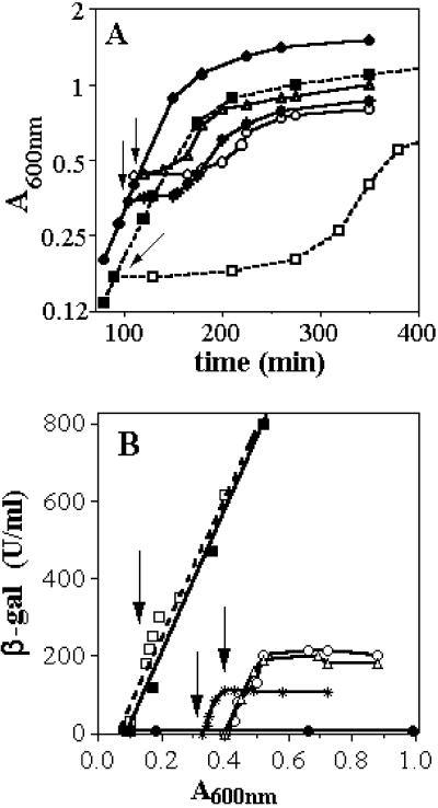
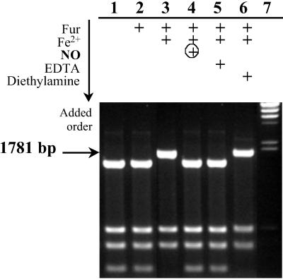
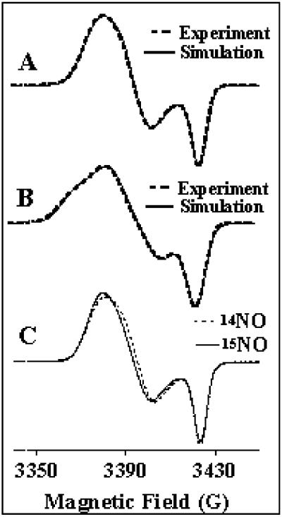
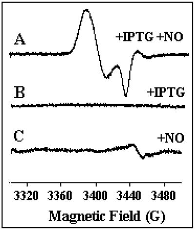
Ferric uptake regulation protein (Fur) is a bacterial global regulator that uses iron as a cofactor to bind to specific DNA sequences. The function of Fur is not limited to iron homeostasis. A wide variety of genes involved in various mechanisms such as oxidative and acid stresses are under Fur control. Flavohemoglobin (Hmp) is an NO-detoxifying enzyme induced by NO and nitrosothiol compounds. Fur recently was found to regulate hmp in Salmonella typhimurium, and in Escherichia coli, the iron-chelating agent 2,2'-dipyridyl induces hmp expression. We now establish direct inhibition of E. coli Fur activity by NO. By using chromosomal Fur-regulated lacZ reporter fusion in E. coli, Fur activity is switched off by NO at micromolar concentration. In vitro Fur DNA-binding activity, as measured by protection of restriction site in aerobactin promoter, is directly sensitive to NO. NO reacts with Fe(II) in purified FeFur protein to form a S = 12 low-spin FeFur-NO complex with a g = 2.03 EPR signal. Appearance of the same EPR signal in NO-treated cells links nitrosylation of the iron with Fur inhibition. The nitrosylated Fur protein is still a dimer and is stable in anaerobiosis but slowly decays in air. This inhibition probably arises from a conformational switch, leading to an inactive dimeric protein. These data establish a link between control of iron metabolism and the response to NO effects.


Figures

Fig 1.
Fig 1.
Effect of NO on growth and β-gal expression in strains carrying fhuF:lacZ fusion. Strains fhuF:lacZ and Δ-Fur fhuF:lacZ were grown in the presence or absence of DEANO in anaerobiosis at 37°C (A) and assayed for β-gal (B). The differential rate of β-gal synthesis is represented as the total β-gal synthesized by milliliter of culture: β-gal activity expressed in Miller units × A600 (β-gal units/ml) in function of the absorbance at 600 nm (A600). Arrows indicate time of addition of DEANO. ○, Strains fhuF:lacZ with 25 μM DEANO; ▵ and *, with 10 μM DEANO; and •, no DEANO; □, Δ-Fur fhuF:lacZ with 200 μM DEANO; ▪, no DEANO.
Fig 2.
Fig 2.
In vitro assay of NO effect on Fur DNA binding. Fifty nanomolar plasmid pDT10 was cleaved by HinfI in the absence or presence of active Fur and after NO treatment. Reaction mixtures were analyzed on 1.5% agarose gel electrophoresis. Lanes: 1, no addition; 2, 20 μM apoFur; 3, 20 μM FeFur; 4, 20 μM FeFur + 40 μM DEANO after 1 h at 20°C; 5, 20 μM FeFur + 50 μM EDTA; 6, 20 μM FeFur + 40 μM diethylamine after 1 h at 20°C; 7, ladder λ DNA HindIII digest (arrow indicates the restriction fragment (1,781 bp) carrying the Fur box that was not cleaved (into 1,530-bp + 251-bp fragments) in the presence of active Fur.
Fig 3.
Fig 3.
EPR spectrum of isotopically labeled FurFe–NO complex. (A) FurFe–NO at 2.5 mM. The simulation is obtained with the g values (2.042; 2.032; 2.015) by using the linewidths 7.9, 7.0, and 4.0 G. (B) Fur57Fe-NO at 3.25 mM. The simulation is achieved by using the [g] values obtained previously with FurFe-NO and the hfs constants A/h (45.6, 35.9, and 3.8 MHz) with the respective linewidths (8.3, 7.5, and 4.6 G). (C) Comparison of Fe(II)-Fur–NO complex at 1.5 mM generated with unlabeled NO and 15NO. The simulations are achieved with the same set of g values (2.042; 2.032; 2.015) and the same linewidths as in A for unlabeled species, but with the respective linewidths (6.7, 7.2, and 3.5 G) for 15NO complex. Nonsaturating EPR conditions: microwave frequency, 9.655 GHz; power, 5 μW; modulation amplitude, 4 G; modulation frequency, 100 kHz; temperature, 30 K.
Fig 4.
Fig 4.
EPR spectrum of intact cells treated with NO. Bacterial strains containing pFur1 were grown at 37°C in aerobiosis from the same preculture and sampled at the same time. (A) Fur synthesis was induced by isopropyl β-d-thiogalactoside during 2 h and 30 min and a sample was taken 60 min after the addition of DEANO (10 μM). (B) A sample was taken after Fur synthesis was induced during 3 h and 30 min without any addition of DEANO. (C) A sample was taken 60 min after the addition of DEANO (10 μM) in noninduced culture. EPR conditions: microwave frequency, 9.655 GHz; power, 5 μW; modulation amplitude, 10 G; modulation frequency, 100 kHz; temperature, 30 K.

References

    1. Neilands J. B. (1972) Struct. Bonding (Berlin) 11, 145-170.
    1. Hibbs J. B., Taintor, R. R., Vavrin, Z. & Rachlin, E. M. (1988) Biochem. Biophys. Res. Commun. 157, 87-94. - PubMed
    1. Marletta M. A., Yoon, P. S., Iyengar, R., Leaf, C. D. & Wishnok, J. S. (1988) Biochemistry 27, 8706-8711. - PubMed
    1. Sari M.-A., Moali, C., Boucher, J. L., Jaouen, M. & Mansuy, D. (1998) Biochem. Biophys. Res. Commun. 250, 364-368. - PubMed
    1. Adak S., Aulak, K. S. & Stuehr, D. J. (2002) J. Biol. Chem. 277, 16167-16171. - PubMed

MeSH terms