. 2002 Dec 24;99(26):16551-5.
doi: 10.1073/pnas.252644499. Epub 2002 Dec 10.

Noninvasive real-time imaging of apoptosis

Affiliations

Noninvasive real-time imaging of apoptosis

Bharathi Laxman et al. Proc Natl Acad Sci U S A. .

Abstract

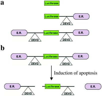
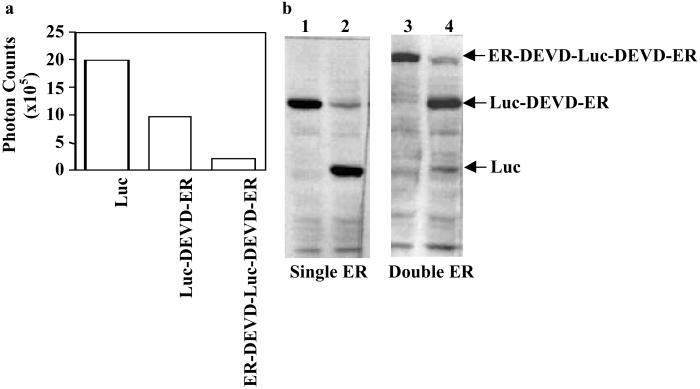
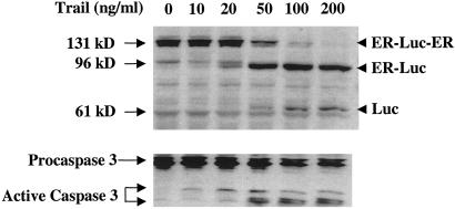
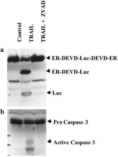
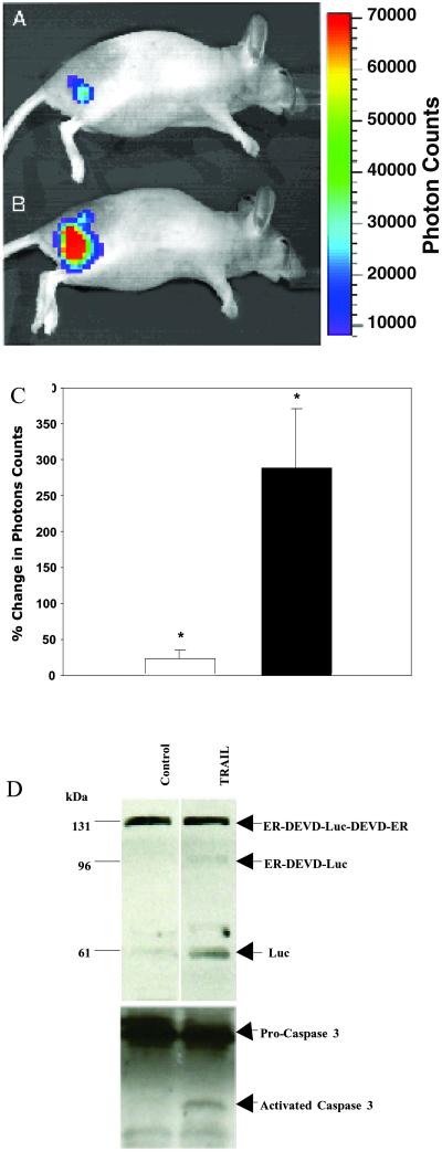
Strict coordination of proliferation and programmed cell death (apoptosis) is essential for normal physiology. An imbalance in these two opposing processes results in various diseases including AIDS, neurodegenerative disorders, myelodysplastic syndromes, ischemiareperfusion injury, cancer, autoimmune disease, among others. Objective and quantitative noninvasive imaging of apoptosis would be a significant advance for rapid and dynamic screening as well as validation of experimental therapeutic agents. Here, we report the development of a recombinant luciferase reporter molecule that when expressed in mammalian cells has attenuated levels of reporter activity. In cells undergoing apoptosis, a caspase-3-specific cleavage of the recombinant product occurs, resulting in the restoration of luciferase activity that can be detected in living animals with bioluminescence imaging. The ability to image apoptosis noninvasively and dynamically over time provides an opportunity for high-throughput screening of proapoptotic and antiapoptotic compounds and for target validation in vivo in both cell lines and transgenic animals.


Figures

Fig 1.
Fig 1.
The strategy for imaging of apoptosis. (a) Cellular expression of a recombinant cDNA encoding reporter molecules was investigated. Shown are the control Luc molecule, Luc fused to a carboxyl-terminal ER, and finally a Luc molecule with ERs at the amino and carboxyl termini. (b) Chimeric polypeptides consisting of a reporter molecule fused to the ER resulted in the most efficient silencing of the reporter activity. Inclusion of a protease cleavage site between these domains provided for protease-mediated activation of the reporter molecule after separation of the silencing domains (i.e., ER). For example, inclusion of the DEVD sequence (a caspase-3 cleavage site) between ER and Luc results in the release of Luc from the silencing domain from the amino and the carboxyl termini in a caspase-3-dependent manner. Because most cells activate caspase-3 during apoptosis, this reporter construct can be used for reporting (imaging) of apoptosis. The cleavage site of this molecular construct can be modified to “report” on the activation of a variety of proteases for detection of proteolytic activity in both cells and transgenic animals by using noninvasive imaging methodologies.
Fig 2.
Fig 2.
In vitro demonstration of the silencing and apoptotic-induced activation of the reporter molecule. (a) Photon counts obtained from cultured D54 glioma cells stably expressing containing Luc, Luc-DEVD-ER, and ER-DEVD-Luc-DEVD-ER reporter molecules were acquired by using the BLI system. The single and double ER fusion molecules were found to attenuate the signal from Luc by ≈50% and 90%, respectively, as compared with native Luc. (b) A Western blot obtained by using a Luc-specific antibody demonstrates that, in response to an apoptosis-inducing agent (TRAIL, lanes 2 and 4), both the Luc-DEVD-ER (lanes 1 and 2) and the ER-DEVD-Luc-DEVD-ER constructs (lanes 3 and 4) were cleaved to yield the Luc protein (61 kDa). Also present was an intermediate cleavage fragment of the two ER-containing molecules to the one ER-containing molecule (lane 4, 96 kDa).
Fig 3.
Fig 3.
Dose-dependent induction of apoptosis in D45 cells expressing the ER-DEVD-Luc-DEVD-ER reporter construct revealed a correlation of caspase-3 activation with cleavage of Luc from the ER silencing domains. (Upper) Using a Luc-specific antibody, the ER-DEVD-Luc-DEVD-ER molecule (131 kDa) is cleaved to a 96-kDa molecule (ER-DEVD-Luc or Luc-DEVD-ER) and subsequently to Luc (61 kDa) when apoptosis is occurring. (Lower) The cleavage event correlated with the conversion of inactive zymogen caspase-3 (32 kDa) to active caspase-3 (17 and 13 kDa).
Fig 4.
Fig 4.
In vitro demonstration of the specificity of the reporter molecule in intact cells. Cultured D54 cells expressing the ER-DEVD-Luc-DEVD-ER reporter molecule were treated with TRAIL and TRAIL + ZVAD, a specific inhibitor of caspase-3. (a) A Western blot was obtained on cell extracts by using a Luc-specific antibody, which demonstrated inhibition of cleavage could be accomplished by inhibition of caspase-3. (b) Cleavage of the reporter molecule during apoptosis was also found to correlate with the conversion of inactive zymogen caspase (32 kDa) to active caspase-3 (17 kDa and 13 kDa).
Fig 5.
Fig 5.
In vivo imaging of caspase-3 activation. In vivo bioluminescence image of a mouse with a s.c. D54-LucER glioma obtained before (A) and 60–75 min after (B) TRAIL treatment. Note the significant increase in photons collected from the same animal after TRAIL treatment. (C) Average percentage change in photon output in PBS-treated (open bar, 21 ± 28%) and TRAIL-treated (filled bar, 287 ± 186% P < 0.04) D54-LucER glioma tumors (n = 5 animals per group) that revealed an ≈3-fold mean increase in bioluminescence on apoptosis induction. Error bars represent SD. (D) Western blot analysis of PBS- and TRAIL-treated excised tumors using antibodies specific for Luc or caspase-3 revealed activation of caspase-3 in TRAIL-treated tumors but not in control (PBS-treated) tumors. A concomitant cleavage (activation) of the 131-kDa reporter was observed in TRAIL-treated tumors.

Similar articles

Cited by

References

    1. Gambhir S. S., Herschman, H. R., Cherry, S. R., Barrio, J. R., Satyamurthy, N., Toyokuni, T., Phelps, M. E., Larson, S. M., Balatoni, J., Finn, R., et al. (2000) Neoplasia 2, 118-138. - PMC - PubMed
    1. Weissleder R., Moore, A., Mahmood, U., Bhorade, R., Benveniste, H., Chiocca, E. A. & Basilion, J. P. (2000) Nat. Med. 6, 351-355. - PubMed
    1. Ponomarev V., Doubrovin, M., Lyddane, C., Beresten, T., Balatoni, J., Bornman, W., Finn, R., Akhurst, T., Larson, S., Blasberg, R., et al. (2001) Neoplasia 3, 480-488. - PMC - PubMed
    1. Doubrovin M., Ponomarev, V., Beresten, T., Balatoni, J., Bornmann, W., Finn, R., Humm, J., Larson, S., Sadelain, M., Blasberg, R. & Tjuvajev, J. G. (2001) Proc. Natl. Acad. Sci. USA 98, 9300-9305. - PMC - PubMed
    1. Rehemtulla A., Hall, D. E., Stegman, L. D., Chen, G., Bhojani, M. S., Chenevert, T. L. & Ross, B. D. (2002) Mol. Imaging 1, 43-55. - PubMed

Publication types