. 2002 Dec 24;99(26):16654-9.
doi: 10.1073/pnas.262591699. Epub 2002 Dec 10.

Excision of misincorporated ribonucleotides in DNA by RNase H (type 2) and FEN-1 in cell-free extracts

Affiliations

Excision of misincorporated ribonucleotides in DNA by RNase H (type 2) and FEN-1 in cell-free extracts

Bjorn Rydberg et al. Proc Natl Acad Sci U S A. .

Abstract

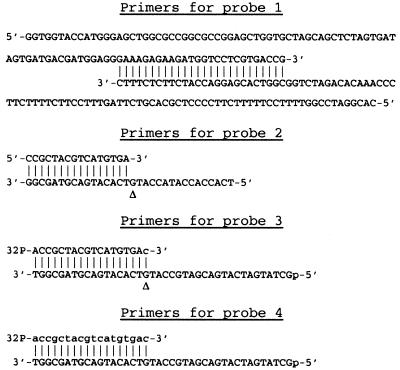
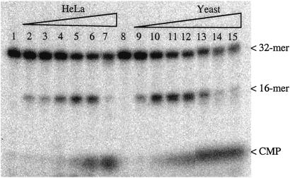
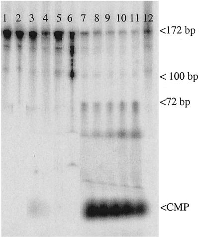
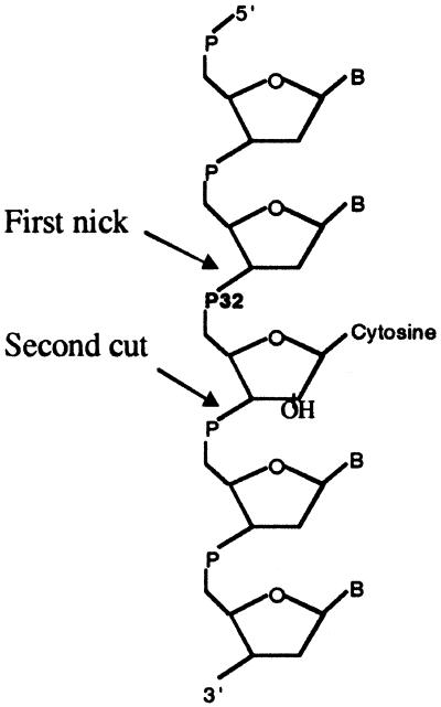
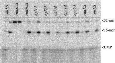
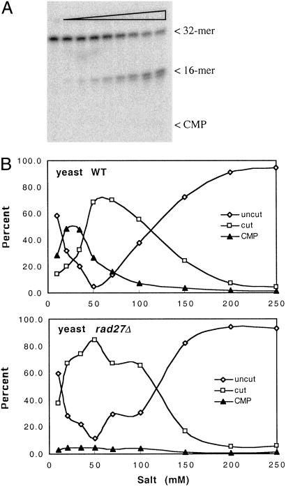
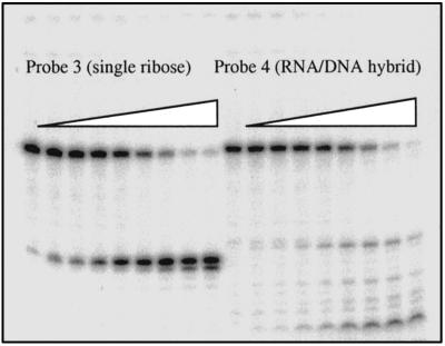
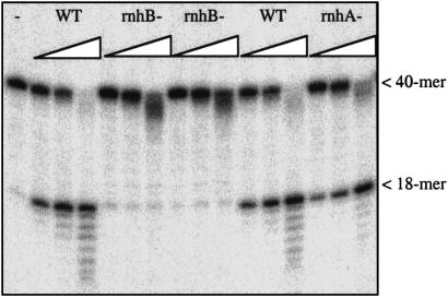
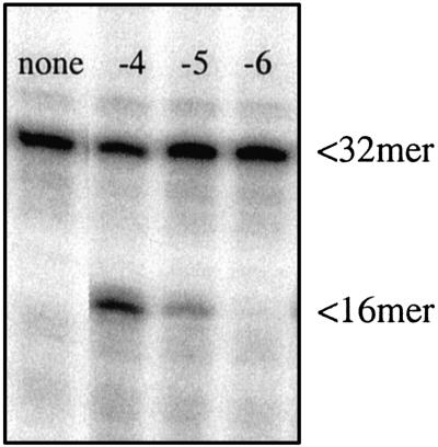
Misincorporated ribonucleotides in DNA will cause DNA backbone distortion and may be targeted by DNA repair enzymes. Using double-stranded oligonucleotide probes containing a single ribose, we demonstrate a robust activity in human, yeast, and Escherichia coli cell-free extracts that nicks 5' of the ribose. The human and yeast extracts also make a subsequent cut 3' of the ribonucleotide releasing a ribonucleotide monophosphate. The resulting 1-nt gap is an ideal substrate for polymerase and ligase to complete a proposed repair sequence that effectively replaces the ribose with deoxyribose. Screening of yeast deletion mutant cells reveals that the initial nick is made by RNase H(35), a RNase H type 2 enzyme, and the second cut is made by Rad27p, the yeast homologue of human FEN-1 protein. RNase H type 2 enzymes are present in all kingdoms of life and are evolutionarily well conserved. We knocked out the corresponding rnhb gene in E. coli and show that extracts from this strain lack the nicking activity. Conversely, a highly purified archaeal RNase HII type 2 protein has a pronounced activity. To study substrate specificity, extracts were made from a yeast double mutant lacking the other main RNase H enzymes [RNase H1 and RNase H(70)], while maintaining RNase H(35). It was found that a single ribose is preferred as substrate over a stretch of riboses, further strengthening a proposed role of this enzyme in the repair of misincorporated ribonucleotides rather than (or in addition to) processing RNADNA hybrid molecules.


Figures

Fig 1.
Fig 1.
Primers used for construction of probes containing a single ribose residue (probes 1–3) or a stretch of ribose residues (probe 4). Capital letters are used for deoxyribonucleotides and lowercase letters are used for ribonucleotides. A triangle (Δ) indicates the position of ribose misincorporation for probe 2 and the location of ribose residue in probe 3.
Fig 2.
Fig 2.
Nicking of probe 2 by crude cell-free extracts. Lanes 1 and 8, no extract; lanes 2–7, increasing amounts of HeLa cell extract (0.001–0.1 μl/assay); lanes 9–15, increasing amounts of S. cerevisiae extract (0.004–0.5 μl/assay).
Fig 3.
Fig 3.
Nicking of probe 1. Lanes 1–4, control probe with no ribose incorporation; lanes 5–12, with ribo C incorporation; lanes 1 and 5, no extract; lanes 2 and 6, probe treated with KOH; lanes 3 and 7–11, incubation with various amounts of Jurkat crude extract; lanes 4 and 12, incubation with heat-inactivated extract.
Fig 4.
Fig 4.
Excision of ribonucleotide from oligomer probes by crude cell-free extracts.
Fig 5.
Fig 5.
Crude extracts from 10 S. cerevisiae knockout strains were assayed for nicking activity by using probe 2. Each strain was assayed with two extract concentrations (1 and 0.1 μl per assay). RNH1, RNH35, and RNH70 are the genes for the three known RNase H type enzymes present in S. cerevisiae. NGL1, NGL2, NGL3, and YEN1 are putative endonuclease genes, as determined by sequence homology. APN1 and APN2 are AP-endonuclease genes, and RAD1 is a gene for a 5′-endonuclease that functions in nucleotide excision repair. Note the absence of nicking in the extract generated from the rnh35Δ knockout strain. The amount of protein in the extracts was in the range of 1.4–2.1 mg/ml.
Fig 6.
Fig 6.
(A) Increasing amounts of extract from a rad27Δ knockout cell line were incubated with probe 2. Compared with extracts with wild-type RAD27 (Fig. 5), only a very limited amount of CMP is seen, with the majority of product being the 16 mer even at a high concentration of extract per assay. (B) Products generated as a function of salt concentration in the assay for wild-type (Upper) and rad27Δ knockout (Lower) cell lines. Note the absence of CMP in the reaction using extract from the rad27Δ mutant.
Fig 7.
Fig 7.
Probes 3 and 4 were incubated with increasing amounts of crude extract from a double yeast mutant lacking RNaseH1 and RnaseH(70). The single ribose in probe 3 is the preferred substrate.
Fig 8.
Fig 8.
Nicking of probe 3 (40 mer, leftmost lane) by increasing amounts of E. coli extracts from wild-type strain (WT), two independent rnhB knockout strains, and a rnhA− strain. Laddering is due to nonspecific nucleases in the extracts. Note the lack of specific nicking for the rnhB knockouts.
Fig 9.
Fig 9.
Nicking of probe 2 by purified P. furiosus RNase HII. First lane: no enzyme. Next three lanes: 1 μl of enzyme added after dilution of the original stock solution 10−4 to 10−6, as indicated. The original stock solution had a concentration of 50 μg/μl.

References

    1. von Sonntag C. & Schulte-Frohlinde, D. (1978) Mol. Biol. Biochem. Biophys. 27, 204-226. - PubMed
    1. Isildar M., Schuchmann, M. N., Schulte-Frohlinde, D. & von Sonntag, C. (1981) Int. J. Radiat. Biol. 40, 347-354. - PubMed
    1. von Sonntag C. (1984) Int. J. Radiat. Biol. 46, 507-519. - PubMed
    1. Joyce C. M. (1997) Proc. Natl. Acad. Sci. USA 94, 1619-1622. - PMC - PubMed
    1. Horton N. C. & Finzel, B. C. (1996) J. Mol. Biol. 264, 521-533. - PubMed

Publication types

LinkOut - more resources