. 2002 Dec 24;99(26):16642-7.
doi: 10.1073/pnas.262659699. Epub 2002 Dec 10.

The structure of Escherichia coli BtuF and binding to its cognate ATP binding cassette transporter

Affiliations

The structure of Escherichia coli BtuF and binding to its cognate ATP binding cassette transporter

Elizabeth L Borths et al. Proc Natl Acad Sci U S A. .

Abstract

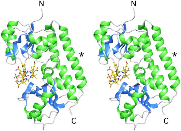
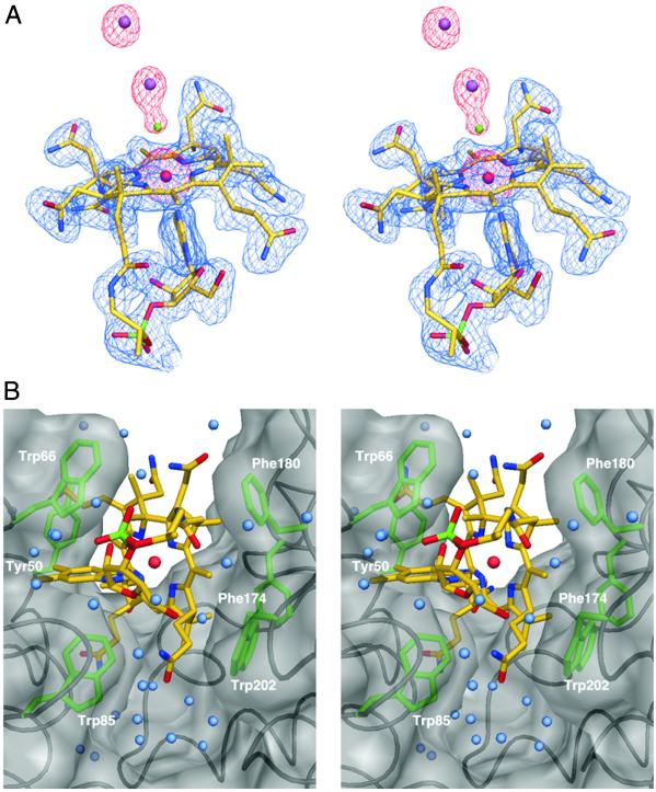
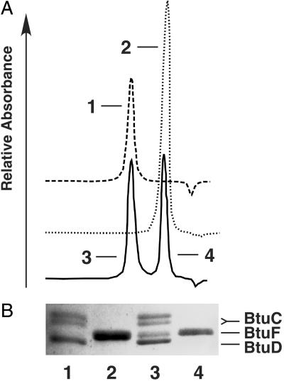
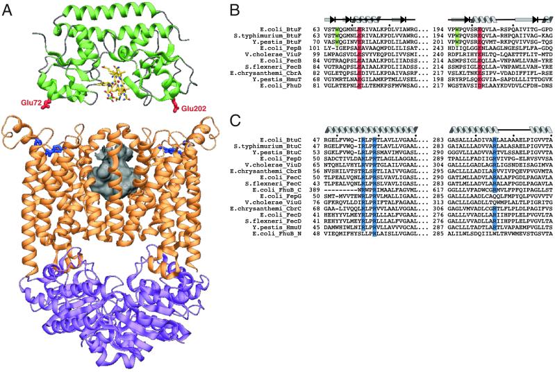
Bacterial binding protein-dependent ATP binding cassette (ABC) transporters facilitate uptake of essential nutrients. The crystal structure of Escherichia coli BtuF, the protein that binds vitamin B12 and delivers it to the periplasmic surface of the ABC transporter BtuCD, reveals a bi-lobed fold resembling that of the ferrichrome binding protein FhuD. B12 is bound in the "base-on" conformation in a deep cleft formed at the interface between the two lobes of BtuF. A stable complex between BtuF and BtuCD (with the stoichiometry BtuC2D2F) is demonstrated to form in vitro and was modeled using the individual crystal structures. Two surface glutamates from BtuF may interact with arginine residues on the periplasmic surface of the BtuCD transporter. These glutamate and arginine residues are conserved among binding proteins and ABC transporters mediating iron and B12 uptake, suggesting that they may have a role in docking and the transmission of conformational changes.


Figures

Fig. 1.
Fig. 1.
Structure of BtuF. A ribbon diagram of BtuF is shown in stereo with bound vitamin B12 in ball and stick. The N and C termini are labeled N and C, respectively. The backbone α-helix that bridges the two lobes of the protein is marked with an asterisk (see text).
Fig. 2.
Fig. 2.
Stereo view of vitamin B12 bound to BtuF. (A) B12 is shown in ball and stick with carbon atoms in yellow, oxygens in red, nitrogens in blue, and phosphorus in green. The central cobalt atom is depicted as a red sphere. The experimental electron density at 2-Å resolution is shown as a blue mesh contoured at 1.2 σ, whereas the anomalous difference Fourier density is shown as a red mesh and contoured at 8 σ. Cadmium and chlorine ions are shown as purple and green spheres, respectively. (B) B12 binding site. The BtuF backbone is shown in black and the molecular surface of BtuF in transparent gray. Side chains of six aromatic residues in van der Waals contact with B12 are colored green. B12 is shown as in A, whereas water molecules are depicted as light blue spheres.
Fig. 3.
Fig. 3.
In vitro interaction of BtuF and BtuCD. (A) BtuF and BtuCD were mixed in detergent solution (0.1% lauryldimethylamine-N-oxide) at a molar ratio of ≈5:1 in the presence of vitamin B12. The mixture was loaded onto an Superdex 200 10/30 column (Amersham Pharmacia) with pure BtuF (dotted line) and BtuCD (dashed line) as controls. Note that because of the presence of a large detergent micelle, pure BtuCD elutes at essentially the same volume as the complex of BtuCD and BtuF. (B) Peak fractions from the gel filtration chromatography shown in A were analyzed by SDS/PAGE. Lane numbers correspond to peaks as labeled in A. Note that BtuC runs as a doublet.
Fig. 4.
Fig. 4.
Proposed interaction between BtuF and BtuCD. (A) BtuF and BtuCD are depicted as ribbon diagrams in an orientation that places the BtuF surface glutamates adjacent to conserved BtuC arginines (see text for further explanation). Once docked, the molecules were separated along the vertical axis for clarity. BtuF is shown in green, with the critical glutamates shown in ball and stick and colored red. BtuC and BtuD are shown in orange and purple, respectively, with the critical BtuC arginines (Arg-56, -59, and -295) shown in ball and stick and colored blue. The size and location of the translocation pathway at the interface of the BtuC subunits (8) are represented by the molecular surface colored in gray. (B) Sequence alignment of iron siderophore/cobalamin binding proteins. The conserved surface glutamates are shown with a red background, with tryptophans contacting B12 on the green background. The secondary structure elements assigned for the E. coli BtuF structure are indicated above the sequence. (C) Alignment of the membrane-spanning subunits of the cognate ABC transporters with conserved arginines shown with a blue background. The secondary structure elements assigned for the structure of E. coli BtuC (8) are indicated above the sequence.

References

    1. Higgins C. F. (2001) Res. Microbiol. 152, 205-210. - PubMed
    1. Dean M., Rzhetsky, A. & Allikmets, R. (2001) Gen. Res. 11, 1156-1166. - PubMed
    1. Nikaido H. & Hall, J. A. (1998) Methods Enzymol. 292, 3-20. - PubMed
    1. Boos W. & Lucht, J. M. (1996) in Escherichia coli and Salmonella typhimurium: Cellular and Molecular Biology, ed. Neidhardt, F. C. (Am. Soc. Microbiol., Washington, DC), Vol. 1, pp. 1175–1209.
    1. Davidson A. L. (2002) J. Bacteriol. 184, 1225-1233. - PMC - PubMed

MeSH terms