. 2002 Dec;13(12):4179-94.
doi: 10.1091/mbc.01-07-0373.

Regulation of IkappaBbeta expression in testis

Affiliations

Regulation of IkappaBbeta expression in testis

Lucy M Budde et al. Mol Biol Cell. 2002 Dec.

Abstract

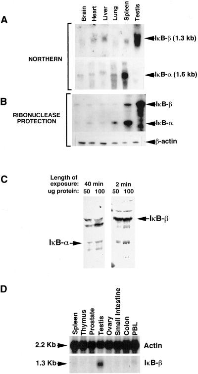
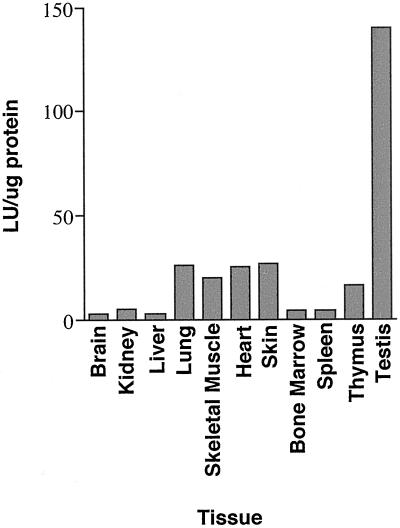
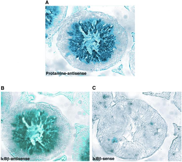
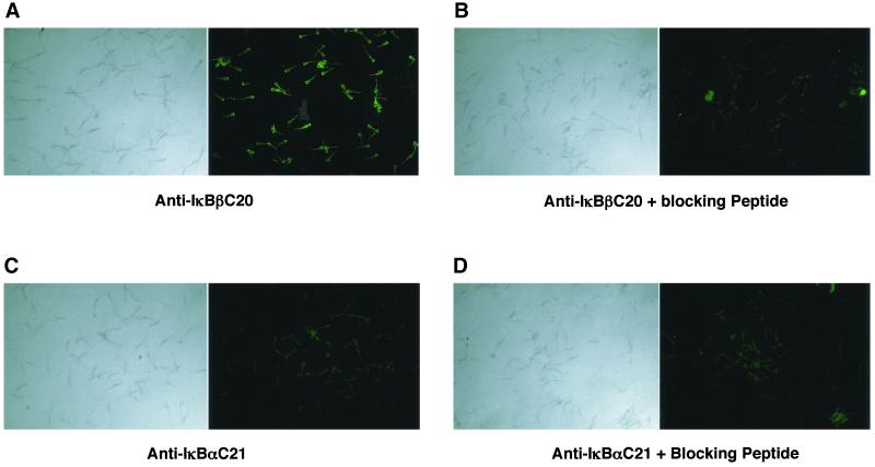
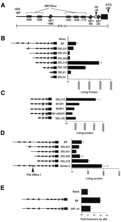
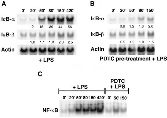
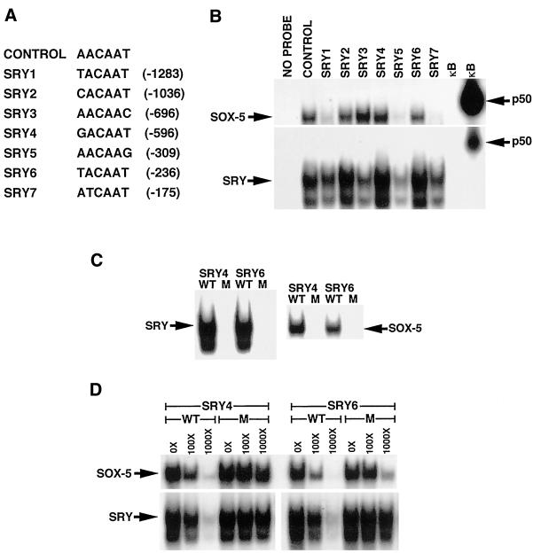
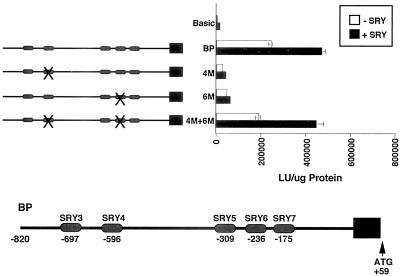
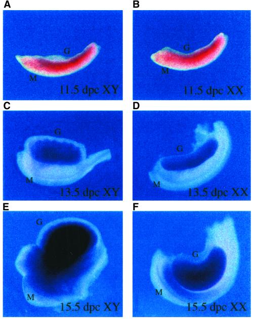
IkappaBalpha and IkappaBbeta are regulators of the nuclear factor-kappaB (NF-kappaB) transcription factor family. Both IkappaBs bind to the same NF-kappaB dimers and are widely expressed in different cells and tissues. To better understand how these two IkappaB isoforms differ biologically, we have characterized the expression of IkappaBbeta in testis, a tissue in which IkappaBalpha is only minimally expressed. We have found that IkappaBbeta expression is localized within the haploid spermatid stages of spermatogenesis and follows the expression of nuclear NF-kappaB. IkappaBbeta expression in haploid spermatids is likely regulated by Sox family proteins, members of which are also expressed within spermatids. We have shown that both SRY and Sox-5 can bind to multiple Sox binding sites found within the IkappaBbeta promoter and can enhance transcription of a reporter gene in transient transfection assays. We also demonstrate that IkappaBbeta mRNA is strongly expressed in developing male gonads. These results therefore suggest that IkappaBbeta may be a novel target for transcription factors of the HMG-box SRY/Sox family and imply a potential role for NF-kappaB/IkappaBbeta in spermatogenesis.


Figures

Figure 1
Figure 1
IκBβ but not IκBα is highly expressed in mouse testes. Multiple tissue Northern blot (A) and ribonuclease protection (B) analyses show that IκBβ mRNA is highly expressed within the testis more than in any other tissue tested. In contrast, IκBα mRNA is barely detectable within the testis. (C) Western blot analysis of total testis extracts shows that this difference in IκBβ and IκBα expression holds up at the protein level. The left panel was probed with an IκBα antibody and the right panel was probed with an IκBβ antibody. (D) Northern blot analysis of a multiple tissue blot shows that high level IκBβ expression does not occur in ovary, suggesting that this expression is not a general phenomenon of reproductive tissues.
Figure 2
Figure 2
Tissue distribution of luciferase activity in mice containing an NF-κB–dependent reporter transgene. Total cell extracts were made from the tissues indicated and assayed for luciferase reporter gene activity. The data shown are representative of results obtained in repeated experiments.
Figure 3
Figure 3
Expression of IκBβ mRNA in mouse testes occurs in haploid spermatids. Tissue sections of adult mouse testes were hybridized to antisense protamine (A), antisense IκBβ (B), or sense IκBβ (C) probes. Similar to the staining seen for protamine mRNA, staining for IκBβ mRNA occurs within the haploid spermatids, located toward the lumen of the seminiferous tubules. As expected, no staining is seen with the IκBβ sense probe.
Figure 4
Figure 4
Immunostaining for IκBβ in bull sperm. (A) Ejaculated bull sperm were stained with antibody against IκBβ protein and visualized with FITC-conjugated secondary antibody. (B) IκBβ antibody was preincubated with the blocking peptide before use. (C) Sperm were stained with antibody against IκBα protein and visualized with a FITC-conjugated secondary antibody. (D) IκBβ antibody was preincubated with the blocking peptide before use. Bright field microscopy of sperm shown in the left-hand panels.
Figure 5
Figure 5
Characterization of the IκBβ promoter. (A) Schematic representation of putative binding sites identified in the IκBβ promoter. The initiating methionine (ATG) is located at +59. (B) Boundaries of the IκBβ promoter were established using luciferase reporter constructs in which the 879 nucleotides upstream of the ATG were progressively deleted at the 5′ end. Data shown are from transient transfection of HeLa cells. Maximal reporter activity was observed upon deletion of 503 of the most 5′ nucleotides in the DEL318 construct, suggesting important positive regulatory elements were located upstream and that a negative regulatory element existed upstream. (C) Contribution of the two downstream SP1 sites to the reporter activity observed for the DEL318 construct was established by mutating the SP1 sites individually and together. Both SP1 sites contribute significantly to reporter activity in Jurkat cells. (D) Demonstration that a negative regulatory element is located between nucleotides −449 and −376. Replacement of this sequence in the BP construct with another segment of DNA of equivalent size results in increased reporter activity in Jurkat cells. (E) NF-κB activation by p65 cotransfection in Jurkat cells leads to increased reporter activity of the BP construct through NF-κB binding to the κB site, as mutation of this site inhibits increased reporter activity.
Figure 6
Figure 6
Comparison of IκBβ and IκBα mRNA induction by NF-κB in 70Z/3 cells stimulated with LPS. (A) Total RNA was isolated from cells stimulated with 10 μg/ml LPS for the indicated times and probed with radiolabeled fragments of IκBα, IκBβ, or actin cDNAs after Northern hybridization. Fold induction was determined by normalization to actin signals by densitometric analysis and is indicated below each time point. (B) Cells were pretreated for 1 h with 100 μM PDTC to inhibit the activation of NF-κB by LPS treatment of cells to establish that mRNA induction was due to NF-κB. (C) Gel shift analysis of total cell extracts incubated with a κB probe to establish that NF-κB was activated by LPS and inhibited by PDTC pretreatment in A and B.
Figure 7
Figure 7
SRY and Sox-5 bind to the SRY/Sox protein binding sites in the IκBβ promoter. (A) Sequence of the individual Sox protein binding sites within the IκBβ promoter. The sequence of the previously determined preferred Sox protein binding site used as the positive control is also listed. (B) Purified His-tagged SRY or Sox-5 (100 ng) was incubated with a radiolabeled oligonucleotide of each putative SRY/Sox family binding site and run on a nondenaturing polyacrylamide gel. Oligonucleotides containing the perfect Sox protein consensus and a κB consensus sequence were included as positive and negative controls, respectively. In vitro-translated p50 protein was incubated with the κB oligonucleotide as an additional control. (C) His-tagged SRY or Sox-5 (100 ng) bound to radiolabeled oligonucleotides containing wild-type but not mutant Sox protein binding sites SRY4 and SRY6. (D) Cold oligonucleotides containing wild-type but not mutant Sox protein binding sites SRY4 and SRY6 competed for binding to 100 ng of His-tagged SRY or Sox-5 bound to wild-type radiolabeled oligonucleotides SRY4 and SRY6.
Figure 8
Figure 8
SRY increases activity of an IκBβ promoter luciferase reporter construct. Cotransfection of an SRY-encoding plasmid and the BP reporter construct into HeLa cells results in a twofold increase in reporter activity. Mutation of Sox protein binding sites 4 (4 M) or 6 (6 M) in the BP construct inhibited the increase in reporter activity, whereas mutation of both sites 4 and 6 together (4 M+6 M) did not prevent an increase in reporter activity.
Figure 9
Figure 9
Expression of IκBβ in embryonic mouse gonads. The developing gonadal ridge was dissected from male (A, C, and E) and female (B, D, and F) mouse embryos at the indicated number of days postcoitum (p.c.), sectioned, and in situ hybridization performed using an IκBβ antisense probe. Although IκBβ mRNA is expressed at 11.5 d p.c. in the male gonad (A), it is also expressed in the female gonad at the same time (B), suggesting that IκBβ is not an SRY target gene. IκBβ expression continues in the gonads of both sexes at low levels at day 13.5 p.c. (C and D). Strikingly, a dramatic male-specific increase in expression occurs at day 15.5 p.c. (E), with an apparent localization to the developing testis cords.

References

    1. Andersen B, Pearse RV 2nd, Schlegel PN, Cichon Z, Schonemann MD, Bardin CW, Rosenfeld MG. Sperm 1: a POU-domain gene transiently expressed immediately before meiosis I in the male germ cell. Proc Natl Acad Sci USA. 1993;90:11084–11088. - PMC - PubMed
    1. Arenzana-Seisdedos F, Thompson J, Rodriguez MS, Bachelerie F, Thomas D, Hay RT. Inducible nuclear expression of newly synthesized I kappa B alpha negatively regulates DNA-binding and transcriptional activities of NF-kappa B. Mol Cell Biol. 1995;15:2689–2696. - PMC - PubMed
    1. Bartke A. Apoptosis of male germ cells, a generalized or a cell type-specific phenomenon? Endocrinology. 1995;136:3–4. - PubMed
    1. Beg AA, Baldwin AS., Jr The I kappa B proteins: multifunctional regulators of Rel/NF-kappa B transcription factors. Genes Dev. 1993;7:2064–2070. - PubMed
    1. Beg AA, Sha WC, Bronson RT, Baltimore D. Constitutive NF-kappa B activation, enhanced granulopoiesis, and neonatal lethality in I kappa B alpha-deficient mice. Genes Dev. 1995;9:2736–2746. - PubMed

Publication types

MeSH terms

LinkOut - more resources