. 2002 Dec;13(12):4355-70.
doi: 10.1091/mbc.e02-06-0346.

Ran GTPase cycle and importins alpha and beta are essential for spindle formation and nuclear envelope assembly in living Caenorhabditis elegans embryos

Affiliations

Ran GTPase cycle and importins alpha and beta are essential for spindle formation and nuclear envelope assembly in living Caenorhabditis elegans embryos

Peter Askjaer et al. Mol Biol Cell. 2002 Dec.

Erratum in

  • Mol Biol Cell. 2004 Jan;15(1):preceding Table of Contents

Abstract

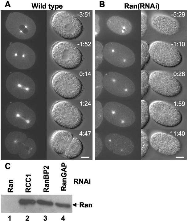
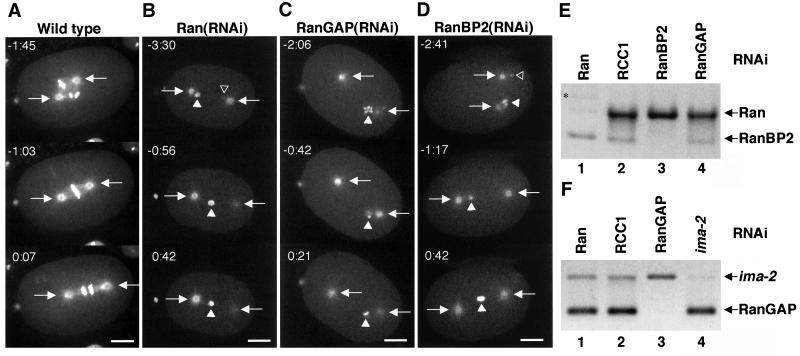
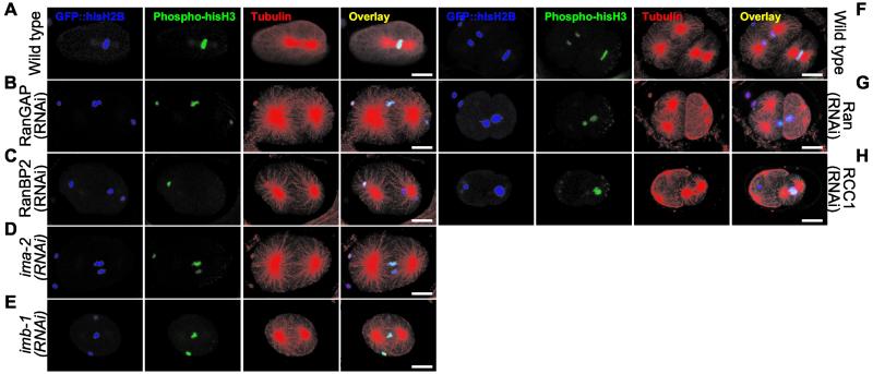
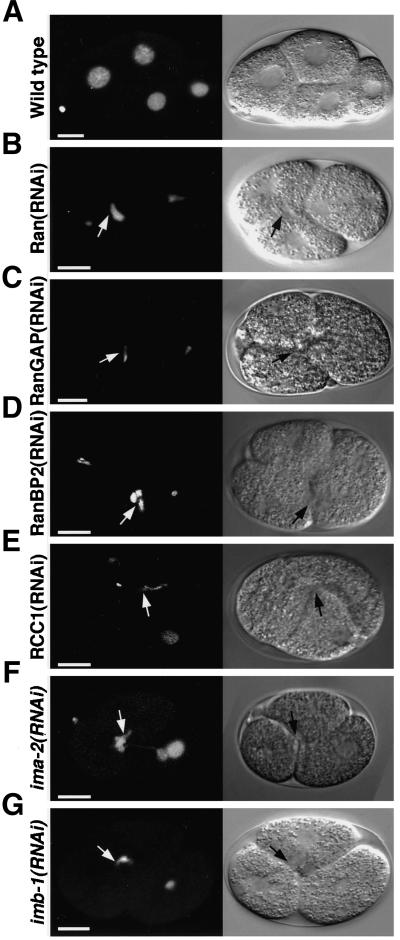
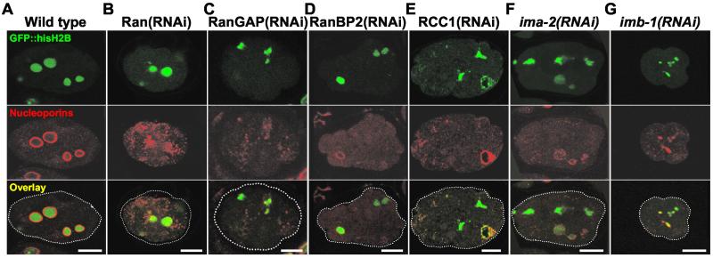
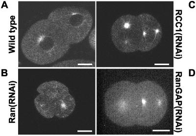
The small GTPase Ran has been found to play pivotal roles in several aspects of cell function. We have investigated the role of the Ran GTPase cycle in spindle formation and nuclear envelope assembly in dividing Caenorhabditis elegans embryos in real time. We found that Ran and its cofactors RanBP2, RanGAP, and RCC1 are all essential for reformation of the nuclear envelope after cell division. Reducing the expression of any of these components of the Ran GTPase cycle by RNAi leads to strong extranuclear clustering of integral nuclear envelope proteins and nucleoporins. Ran, RanBP2, and RanGAP are also required for building a mitotic spindle, whereas astral microtubules are normal in the absence of these proteins. RCC1(RNAi) embryos have similar abnormalities in the initial phase of spindle formation but eventually recover to form a bipolar spindle. Irregular chromatin structures and chromatin bridges due to spindle failure were frequently observed in embryos where the Ran cycle was perturbed. In addition, connection between the centrosomes and the male pronucleus, and thus centrosome positioning, depends upon the Ran cycle components. Finally, we have demonstrated that both IMA-2 and IMB-1, the homologues of vertebrate importin alpha and beta, are essential for both spindle assembly and nuclear formation in early embryos.


Figures

Figure 1
Figure 1
Ran is required for spindle formation. Still images from time-lapse microscopy are shown from wild-type (A) and Ran(RNAi) (B) embryos expressing GFP::α-tubulin. Anterior of the embryos is on the left and time (minutes:seconds) relative to anaphase onset is indicated. For the wild-type embryo the following events are depicted: pronuclear meeting (−3:51), pronuclear envelope breakdown (−1:52), metaphase-to-anaphase transition (0:14), anaphase (1:24), and telophase and cytokinesis (4:47). In the Ran(RNAi) embryo no spindle is formed, nevertheless the embryo eventually divides (11:40). Bar, 10 μm. (C) Western blot analysis of Ran in embryonic lysates from worms depleted of Ran (lane 1), RCC1 (lane 2), RanBP2 (lane 3), or RanGAP (lane 4). Expression of Ran is efficiently and specifically inhibited by Ran dsRNA. Equal loading of protein was verified with antibodies against LEM-2 (our unpublished data).
Figure 2
Figure 2
Depletion of Ran, RanGAP, or RanBP2 causes similar defects on spindle formation. Embryos expressing GFP::β-tubulin and GFP::histone H2B from either control worms (A) or worms depleted of Ran (B), RanGAP (C), or RanBP2 (D) were observed by time-lapse microscopy. Still images are shown with indication of time (minutes:seconds) relative to anaphase onset. Arrows point to the centrosomes, whereas open and closed triangles indicate the pronucleus originating from the sperm and the oocyte, respectively. The sperm pronucleus is in the focal plane only in certain images. Note that the chromatin aligns on a metaphase plate in the wild-type embryo, whereas the pronuclei fail to meet in RNAi embryos and no spindle is formed. Bar, 10 μm. (E) RNA prepared from embryos depleted of Ran (lane 1), RCC1 (lane 2), RanBP2 (lane 3), or RanGAP (lane 4) was analyzed by RT-PCR with primers specific for Ran and RanBP2. Ran and RanBP2 mRNA are efficiently and specifically depleted by dsRNA corresponding to Ran and RanBP2, respectively. Star in lane 1 indicates a RanBP2 PCR product from contaminating genomic DNA. (F) RNA prepared from embryos depleted of Ran (lane 1), RCC1 (lane 2), RanGAP (lane 3), or IMA-2 (lane 4) was analyzed by RT-PCR with primers specific for RanGAP and ima-2. RanGAP and ima-2 mRNA are efficiently and specifically depleted by dsRNA corresponding to RanGAP and ima-2, respectively. Similarly, RCC1 mRNA was inhibited by RCC1 dsRNA (our unpublished data; Figure 4C).
Figure 3
Figure 3
RNAi embryos fail to assemble microtubules into spindles around mitotic chromatin. Embryos from GFP::histone H2B worms grown on either control bacteria (A and F), or bacteria expressing dsRNA corresponding to RanGAP (B), RanBP2 (C), ima-2 (D), imb-1 (E), Ran (G), or RCC1 (H) were fixed and analyzed by immunofluorescence. GFP::histone H2B (depicted in blue) was used to visualize total chromatin, whereas mitotic chromatin and microtubules were recognized with anti-phospho-histone H3 (green) and anti-α-tubulin (red) antibodies. An overlay of the three stains is shown on the right. Bar, 10 μm.
Figure 4
Figure 4
Depletion of RCC1 leads to defects in chromosome segregation. Embryos expressing GFP::β-tubulin and GFP::histone H2B from either control worms (A) or worms depleted of RCC1 (B) were observed by time-lapse microscopy. Still images are shown with indication of time (minutes:seconds) relative to anaphase onset. Anterior of the embryos is on the left. RCC1 depletion causes problems in the initial phase of spindle formation and in DNA segregation. Bar, 10 μm. (C) Comparison of one-cell embryos from GFP::RCC1 worms grown on either control bacteria (left), or bacteria expressing dsRNA corresponding to RCC1 (middle). RNAi against RCC1 decreases the level of fluorescence to that of N2 embryos with no GFP gene (right). Dotted white lines indicate pronuclei and plasma membranes. Bar, 10 μm. (D) Distance between the two centrosomes was measured relative to the total length of the embryo. Shown is the average of several wild-type (diamonds, n = 9), RCC1(RNAi) (squares, n = 10), and RanGAP(RNAi) (triangles, n = 5) embryos as a function of time. Each embryo was aligned relative to anaphase onset, which defines t0. Vertical lines represent the SD at each time point. Also shown is a single Ran(RNAi) embryo (crosses). For wild-type embryos, approximate timing of pronuclear meeting and pronuclear envelope breakdown (NEBD) is indicated as is DNA segregation and spindle rocking.
Figure 5
Figure 5
Embryos devoid of IMA-2 or IMB-1 fail to form mitotic spindles. Embryos expressing GFP::β-tubulin from either a wild-type hermaphrodite (A) or from an ima-2(ok256) homozygous worm (B) were observed by time-lapse microscopy. Still images are shown with indication of time (minutes:seconds) relative to anaphase onset. Bar, 10 μm. (C) Distance between the two centrosomes were measured relative to the total length of the embryo. Shown is the average of several wild-type (diamonds, n = 9; same as in Figure 4C) and ima-2(ok256) (crosses, n = 5) embryos as function of time. Each embryo was aligned relative to anaphase onset, which defines t0. Vertical lines represent the SD at each time point. For wild-type embryos approximate timing of pronuclear meeting and pronuclear envelope breakdown (NEBD) is indicated as is DNA segregation and spindle rocking. (D) An embryo expressing GFP::β-tubulin and GFP::histone H2B from worms depleted of IMB-1 was observed by time-lapse microscopy. Still images are shown with indication of time (minutes:seconds) relative to anaphase onset. Bar, 10 μm. (E) RNA prepared from embryos depleted of RanGAP (lanes 1 and 3) or IMB-1 (lanes 2 and 4) was analyzed by RT-PCR with primers specific for imb-1 (lanes 1 and 2) and Ran (lanes 3 and 4). imb-1 mRNA is efficiently and specifically depleted by dsRNA corresponding to imb-1 (lane 2).
Figure 6
Figure 6
The Ran GTPase cycle, IMA-2, and IMB-1 are essential for normal chromatin appearance. Embryos from GFP::histone H2B worms grown on either control bacteria (A), or bacteria expressing dsRNA corresponding to Ran (B), RanGAP (C), RanBP2 (D), RCC1 (E), ima-2 (F), or imb-1 (G) were observed by time-lapse microscopy. Single still images are shown illustrating that division of P1 is often delayed in RNAi embryos, which leads to prolonged appearance of three-cell-stage embryos. Arrows indicate examples of abnormal chromatin structures that often are trapped at the cleavage furrow. Bar, 10 μm.
Figure 7
Figure 7
RNAi against Ran and its cofactors affects targeting of nuclear pore proteins. Embryos from GFP::histone H2B worms grown on either control bacteria (A), or bacteria expressing dsRNA corresponding to Ran (B), RanGAP (C), RanBP2 (D), RCC1 (E), ima-2 (F), or imb-1 (G) were fixed and analyzed with the anti-nucleoporin mAb mAb414. Top row shows GFP::histone H2B in green, middle row shows mAb414 staining in red, and bottom row shows an overlay of the two signals. Bar, 10 μm.
Figure 8
Figure 8
Disruption of the Ran GTPase cycle prevents nuclear envelope formation. Embryos from GFP::EMR-1 worms grown on either control bacteria (A), or bacteria expressing dsRNA corresponding to Ran (B and C), RanGAP (D), RanBP2 (E), RCC1 (F), ima-2 (G), or imb-1 (H) were observed by time-lapse microscopy. Single still images are shown demonstrating that no nuclear structure surrounded by a uniform EMR-1 staining is seen in embryos where the Ran cycle (B and D–F), IMA-2 (G) or IMB-1 (H) is targeted by RNAi. An example of a less severely affected embryo is shown (C). Bar, 10 μm.
Figure 9
Figure 9
Nuclear exclusion of soluble GFP::β-tubulin depends on the Ran GTPase cycle. Embryos from GFP::β-tubulin worms grown on either control bacteria (A), or bacteria expressing dsRNA corresponding to Ran (B), RCC1 (C), or RanGAP (D) were observed by time-lapse microscopy combined with confocal z-scanning. Single still images are shown illustrating that nuclei in a control embryo exclude GFP::β-tubulin (A), whereas no area of exclusion is detected when the Ran cycle is affected (B–D). Bar, 10 μm.

References

    1. Askjaer P, et al. RanGTP-regulated interactions of CRM1 with nucleoporins and a shuttling DEAD-box helicase. Mol Cell Biol. 1999;19:6276–6285. - PMC - PubMed
    1. Bamba C, Bobinnec Y, Fukuda M, Nishida E. The GTPase Ran regulates chromosome positioning and nuclear envelope assembly in vivo. Curr Biol. 2002;12:503–507. - PubMed
    1. Bilbao-Cortés D, Hetzer M, Laengst G, Becker PB, Mattaj IW. Ran binds to chromatin by two distinct mechanisms. Curr Biol. 2002;12:1151–1156. - PubMed
    1. Bischoff FR, Görlich D. RanBP1 is crucial for the release of RanGTP from importin beta-related nuclear transport factors. FEBS Lett. 1997;419:249–254. - PubMed
    1. Bischoff FR, Ponstingl H. Catalysis of guanine nucleotide exchange on Ran by the mitotic regulator RCC1. Nature. 1991;354:80–82. - PubMed

Publication types

MeSH terms

LinkOut - more resources