

Magnification chromoendoscopy for the detection of intestinal metaplasia and dysplasia in Barrett's oesophagus
- PMID: 12477754
- PMCID: PMC1773525
- DOI: 10.1136/gut.52.1.24
Magnification chromoendoscopy for the detection of intestinal metaplasia and dysplasia in Barrett's oesophagus
Abstract
Background: The presence of intestinal metaplasia (IM) in the columnar lined distal oesophagus defines Barrett's oesophagus with the risk of future malignant transformation. The distribution of both IM and dysplasia (low grade (LGD) and high grade (HGD)) within the columnar lined oesophagus is patchy and mosaic requiring random biopsies. Techniques that could help target areas of high yield within Barrett's mucosa would be helpful.
Aim: To study the utility of high magnification chromoendoscopy (MCE) in the detection of IM, LGD, and HGD in patients with Barrett's oesophagus.
Methods: Consecutive patients detected with columnar mucosa in the distal oesophagus were studied using an Olympus magnification endoscope (GIF-Q16OZ, 115x). The distal oesophagus was sprayed with indigo carmine solution and the oesophageal columnar mucosa patterns were noted under high magnification and targeted for biopsy. All biopsies were read by pathologists blinded to the endoscopic findings.
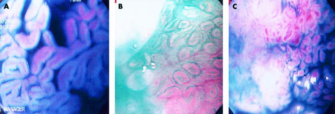
Results: Eighty patients with suspected Barrett's oesophagus (that is, columnar lined distal oesophagus) were studied: mean age 62.7 years (range 35-81). Mean length of columnar mucosa was 3.7 cm (range 0.5-17). Three types of mucosal patterns were noted within the columnar mucosa after spraying indigo carmine and using MCE: ridged/villous pattern, circular pattern, and irregular/distorted pattern. The yield of IM on target biopsies according to the patterns was: ridged/villous 57/62 (97%) and circular 2/12 (17%). Six patients had an irregular/distorted pattern and all had HGD on biopsy (6/6 (100%)). Eighteen patients had LGD on target biopsies; all had the ridged/villous pattern. All patients with long segment Barrett's were identified using MCE whereas 23/28 patients (82%) with short segment Barrett's had the ridged/villous pattern.
Conclusions: MCE helps visually identify areas with IM and HGD having specific patterns but not patients with LGD (appear similar to IM). MCE may be a useful clinical tool for the increased detection of patients with IM as well as for surveillance of patients for the detection of HGD. If these preliminary results are validated, MCE would help identify high yield areas, potentially eliminating the need for random biopsies.
Figures
Comment in
-
[Diagnosis and surveillance in patients with Barrett's esophagus].Z Gastroenterol. 2003 Sep;41(9):939-42. doi: 10.1055/s-2003-41828. Z Gastroenterol. 2003. PMID: 13130333 German. No abstract available.
Comment on
-
S, m, l, xl.Gut. 2003 Jan;52(1):5-7. doi: 10.1136/gut.52.1.5. Gut. 2003. PMID: 12477746 Free PMC article. No abstract available.
References
-
- Weinstein WM, Ippoliti AF. The diagnosis of Barrett’s esophagus: Goblets, goblets, goblets. Gastrointest Endosc 1996;44:91–5. - PubMed
-
- Blot WJ, Devesa SS, Kneller RW, et al. Rising incidence of adenocarcinoma of the esophagus and gastric cardia. JAMA 1991;265:1287–9. - PubMed
-
- Devesa SS, Blot WJ, Fraumeni JF. Changing patterns in the incidence of esophageal and gastric carcinoma in the United States. Cancer 1988;83:2049–53. - PubMed
-
- Hamilton SR, Smith R, Cameron JL. Prevalence and characteristics of Barrett esophagus in patients with adenocarcinoma of the esophagus or esophagogastric junction. Hum Pathol 1988;19:942–8. - PubMed
-
- Sharma P. Endoscopic recognition and screening of Barrett’s esophagus. Techniques Gastrointest Endosc 2000;2:182–5.
Publication types
MeSH terms
Substances
LinkOut - more resources
Full Text Sources
Other Literature Sources
Medical