

Cloning and characterization of scon-3+, a new member of the Neurospora crassa sulfur regulatory system
- PMID: 12477788
- PMCID: PMC138751
- DOI: 10.1128/EC.1.6.875-883.2002
Cloning and characterization of scon-3+, a new member of the Neurospora crassa sulfur regulatory system
Abstract
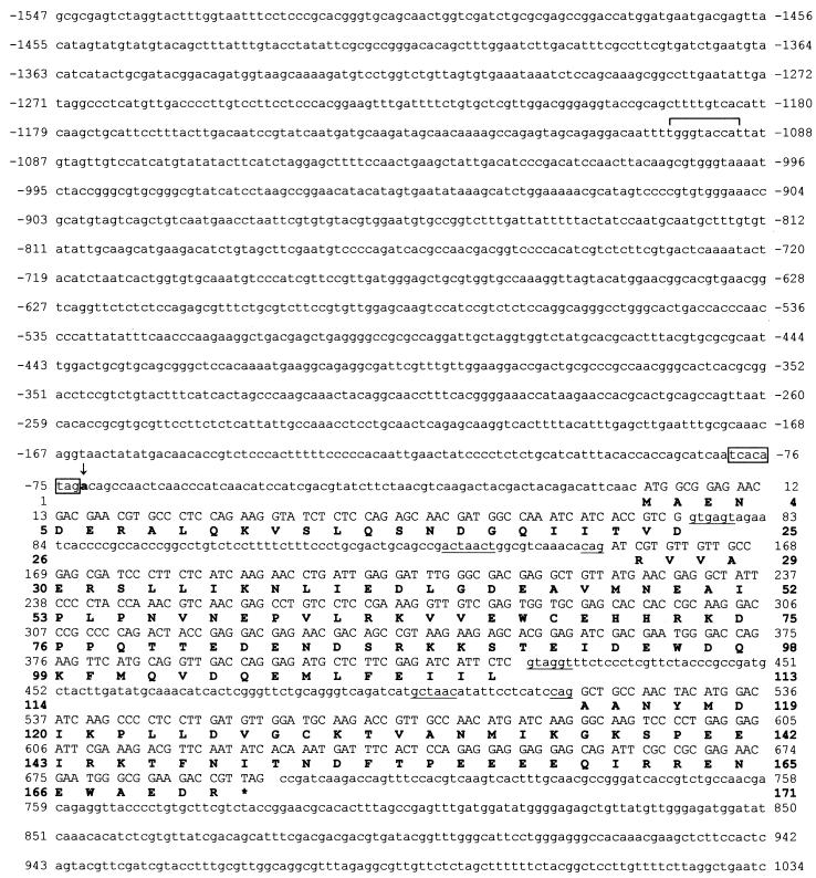
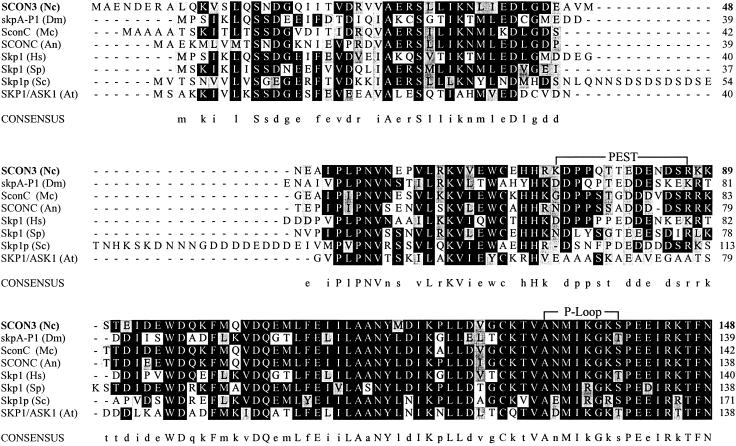
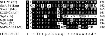
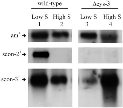
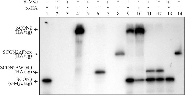
The sulfur regulatory system of Neurospora crassa consists of a group of sulfur-regulated structural genes (e.g., arylsulfatase) that are under coordinate control of the CYS3 positive regulator and sulfur controller (SCON) negative regulators. Here we report on the cloning of scon-3(+), which encodes a polypeptide of 171 amino acids and is a Skp1 family homolog. Repeat-induced point mutation of scon-3(+) resulted in a phenotype of constitutive expression of arylsulfatase, a phenotype consistent with other sulfur controller mutants. Northern analysis indicated that, unlike other members of the sulfur regulatory system, expression of scon-3(+) is not under the direct control of the CYS3 transcriptional activator. In particular, scon-3(+) mRNA was detectable under sulfur repressing or derepressing conditions in a Deltacys-3 mutant. In yeast, Skp1p and an F-box protein binding partner are core constituents of a class of E3 ubiquitin ligases known as SCF complexes. The N. crassa negative regulator SCON2 contains an F-box motif essential for the operation of the sulfur regulatory system and suggests a role for an SCF complex in the N. crassa sulfur regulatory system. A crucial set of experiments, by using a yeast two-hybrid approach with confirming coimmunoprecipitation assays, demonstrated that SCON3 interacts with SCON2 in a manner dependent upon the F-box motif of SCON2. The protein-protein interaction detected between SCON2 and SCON3 represents the initial demonstration in a filamentous fungus of functional interaction between putative core components of a SCF complex.
Figures

Similar articles
-
The sulfur controller-2 negative regulatory gene of Neurospora crassa encodes a protein with beta-transducin repeats.Proc Natl Acad Sci U S A. 1995 Apr 11;92(8):3343-7. doi: 10.1073/pnas.92.8.3343. Proc Natl Acad Sci U S A. 1995. PMID: 7724564 Free PMC article.
-
An additional role for the F-box motif: gene regulation within the Neurospora crassa sulfur control network.Proc Natl Acad Sci U S A. 1998 Mar 3;95(5):2417-22. doi: 10.1073/pnas.95.5.2417. Proc Natl Acad Sci U S A. 1998. PMID: 9482900 Free PMC article.
-
Molecular cloning and analysis of the scon-2 negative regulatory gene of Neurospora crassa.Mol Cell Biol. 1990 Oct;10(10):5207-14. doi: 10.1128/mcb.10.10.5207-5214.1990. Mol Cell Biol. 1990. PMID: 1975945 Free PMC article.
-
Production of the CYS3 regulator, a bZIP DNA-binding protein, is sufficient to induce sulfur gene expression in Neurospora crassa.Mol Cell Biol. 1992 Apr;12(4):1568-77. doi: 10.1128/mcb.12.4.1568-1577.1992. Mol Cell Biol. 1992. PMID: 1532230 Free PMC article.
-
Regulation of sulfur and nitrogen metabolism in filamentous fungi.Annu Rev Microbiol. 1993;47:31-55. doi: 10.1146/annurev.mi.47.100193.000335. Annu Rev Microbiol. 1993. PMID: 8257101 Review.
Cited by
-
Lessons from fungal F-box proteins.Eukaryot Cell. 2009 May;8(5):677-95. doi: 10.1128/EC.00386-08. Epub 2009 Mar 13. Eukaryot Cell. 2009. PMID: 19286981 Free PMC article. Review. No abstract available.
-
The COP9 signalosome regulates the Neurospora circadian clock by controlling the stability of the SCFFWD-1 complex.Genes Dev. 2005 Jul 1;19(13):1518-31. doi: 10.1101/gad.1322205. Epub 2005 Jun 16. Genes Dev. 2005. PMID: 15961524 Free PMC article.
-
Neurospora COP9 signalosome integrity plays major roles for hyphal growth, conidial development, and circadian function.PLoS Genet. 2012;8(5):e1002712. doi: 10.1371/journal.pgen.1002712. Epub 2012 May 10. PLoS Genet. 2012. PMID: 22589747 Free PMC article.
-
Aspects of the Neurospora crassa Sulfur Starvation Response Are Revealed by Transcriptional Profiling and DNA Affinity Purification Sequencing.mSphere. 2021 Oct 27;6(5):e0056421. doi: 10.1128/mSphere.00564-21. Epub 2021 Sep 15. mSphere. 2021. PMID: 34523983 Free PMC article.
-
The Genomes of Three Uneven Siblings: Footprints of the Lifestyles of Three Trichoderma Species.Microbiol Mol Biol Rev. 2016 Feb 10;80(1):205-327. doi: 10.1128/MMBR.00040-15. Print 2016 Mar. Microbiol Mol Biol Rev. 2016. PMID: 26864432 Free PMC article. Review.
References
-
- Bai, C., P. Sen, K. Hofmann, L. Ma, M. Goebl, J. W. Harper, and S. J. Elledge. 1996. SKP1 connects cell cycle regulators to the ubiquitin proteolysis machinery through a novel motif, the F-box. Cell 86:263-274. - PubMed
-
- Bartel, P. L., C.-T. Chien, R. Sternglanz, and S. Fields. 1993. Using the two-hybrid system to detect protein-protein interactions, p. 153-179. In D. A. Hartley (ed.), Cellular interactions in development: a practical approach. Oxford University Press, Oxford, England.
-
- Bruchez, J. J. P., J. Eberle, and V. E. A. Russo. 1993. Regulatory sequences in the transcription of Neurospora crassa genes: CAAT box, TATA box, introns, poly(A) tail formation sequences. Fung. Genet. Newsl. 40:89-96.
-
- Cambareri, E. B., H. M. Foss, M. R. Rountree, and E. U. Selker. 1989. Repeat induced G-C and A-T mutations in Neurospora. Science 244:1571-1575. - PubMed
Publication types
MeSH terms
Substances
LinkOut - more resources
Full Text Sources
Molecular Biology Databases
