. 2002 Dec;1(6):884-94.
doi: 10.1128/EC.1.6.884-894.2002.

The Glc7p-interacting protein Bud14p attenuates polarized growth, pheromone response, and filamentous growth in Saccharomyces cerevisiae

Affiliations

The Glc7p-interacting protein Bud14p attenuates polarized growth, pheromone response, and filamentous growth in Saccharomyces cerevisiae

Paul J Cullen et al. Eukaryot Cell. 2002 Dec.

Abstract

A genetic selection in Saccharomyces cerevisiae for mutants that stimulate the mating pathway uncovered a mutant that had a hyperactive pheromone response pathway and also had hyperpolarized growth. Cloning and segregation analysis demonstrated that BUD14 was the affected gene. Disruption of BUD14 in wild-type cells caused mild stimulation of pheromone response pathway reporters, an increase in sensitivity to mating factor, and a hyperelongated shmoo morphology. The bud14 mutant also had hyperfilamentous growth. Consistent with a role in the control of cell polarity, a Bud14p-green fluorescent protein fusion was localized to sites of polarized growth in the cell. Bud14p shared morphogenetic functions with the Ste20p and Bni1p proteins as well as with the type 1 phosphatase Glc7p. The genetic interactions between BUD14 and GLC7 suggested a role for Glc7p in filamentous growth, and Glc7p was found to have a positive function in filamentous growth in yeast.


Figures

FIG. 1.
FIG. 1.
The bud14 mutant has enhanced pheromone sensitivity and hyperpolarized shmoo morphology. (A) Halo assay. Equal concentrations of wild-type (wt) (SY2002) or bud14 mutant (SY3873) cells were spread onto YPD solid agar medium, and 8 μl of 590 μM alpha factor was applied to a disk at the center of the plate. Plates were incubated for 24 h at 30°C and photographed. (B) Shmoo formation over time. Wild-type (SY2002) and bud14 mutant (SY3873) cells were grown to mid-log phase in YPD medium, washed, and resuspended in YPD medium plus 30 μM alpha factor. Cells were incubated at 30°C, and at the times indicated aliquots were removed and shmoos were scored by microscopic examination. For the y axis, # shmoos refers to the number of shmoos observed in counting 100 cells. (In the bud14 mutant, the number of shmoos exceeds the number of cells because some cells formed multiple shmoos). (C) Shmoo morphology. Wild-type (SY2002), bud14 (SY3873), and GAL-BUD14 mutant (SY3881) cells were grown to mid-log phase, washed, and incubated in 30 μM alpha factor for 3 h at 30°C. Strain SY3881 was grown, washed, and induced in YPGal medium. Cells were photographed at 100×, and representative cells are shown. Bar, 5 μm.
FIG. 2.
FIG. 2.
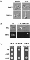
Ste20p and Bud14p share a function in polarized growth. (A) Cell morphology. Cells were grown to mid-log phase in YPD medium for bud14 (SY3873) and bud14 ste20 (SY3876) mutants, or in YPGal for GAL-BUD14 (SY3881) and GAL-BUD14 ste20 (SY3884) mutants. Cells were visualized by microscopy and photographed at 100×. (B) Septin localization. Wild-type (wt) (SY3897) or GAL-BUD14 ste20 (SY3905) cells were grown to mid-log phase in YPGal medium at 30°C. Cells were fixed, permeabilized, and probed with anti-Cdc3p antibodies as described in Materials and Methods. For panels A and B, bar = 5 μm. (C) Ste20p is conditionally required in cells lacking or overproducing Bud14p. Equal concentrations of bud14 (SY3898), bud14 ste20 (SY3908), bud14 ste12 (SY3909), GAL-BUD14 (SY3907), GAL-BUD14 ste20 (SY3905), and GAL-BUD14 ste12 mutant (SY3906) cells were spotted onto the indicated solid agar media and incubated at 30 or 37°C as indicated for 2 days (for SCD and SCD 37°C) or 3 days (for YPGal).
FIG. 3.
FIG. 3.
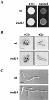
The bud14 mutant exhibits hyperfilamentous growth. (A) Plate-washing assay. Equal concentrations of wild-type (wt) (SY3897) and bud14 mutant (SY3898) cells were spotted onto YPD solid agar medium and incubated for 2 days at 30°C. The plate was photographed (left panel), washed, and photographed again (right panel). (B) Single cell invasive growth assay. Equal concentrations of wild-type (SY3897) and bud14 mutant (SY3898) cells were spread onto SCD (+Glc) or SC (-Glc) medium, incubated for 16 h at 25°C, and photographed at 20×. Bar, 10 μm. (C) Prolonged incubation illustrates the difference in cell length between the wild type (upper panel) and the bud14 mutant (lower panel). Equal concentrations of cells were spotted onto YPD medium and grown for 5 days at 30°C. The plate was washed, and the cells were excised from the plate and photographed at 100×. Bar, 5 μm.
FIG. 4.
FIG. 4.
Contributions of the polarisome and Hsl proteins to the hyperpolarized growth in the bud14 mutant. (A) Combination of bud14 and hsl mutations. Wild-type (wt) (SY3897), bud14 (SY3898), hsl1 (SY3892), hsl7 (SY3893), bud14 hsl1 (SY3894), and bud14 hsl7 mutant (SY3895) cells were grown on YPD solid agar medium for 2 days at 30°C. Cells were removed from the plates, resuspended in water, and photographed at 100×. (B) Mutations that disrupt the polarisome suppress the hyperpolarized growth phenotype conferred by the bud14 mutation. Wild-type (SY3897), bud14 (SY3898), bud14 pea2 (SY3891), bud14 bni1 (SY3890), and bni1 mutant (SY3889) cells were grown on YPD solid agar medium for 2 days at 30°C. Cells were removed from the plates, resuspended in water, and photographed at 100×. For panels A and B, bar = 10 μm. (C) Bni1p is required in cells overexpressing BUD14. Equal concentrations of wild-type (SY3897), bni1 (SY3889), GAL-BUD14 (SY3907), or GAL-BUD14 bni1 (SY3910) mutant cells were spotted onto YPGal solid agar medium and incubated for 2 days at 30°C.
FIG. 5.
FIG. 5.
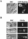
Bud14p localizes to the mother-bud neck and has a role in cytokinesis. (A) GFP-Bud14p localization. Cells containing the galactose-inducible GFP-BUD14 fusion were grown to mid-log phase in YPGal medium at 30°C and examined using DIC (left panel) or a FITC filter (right panel) at 100×. (B) Mislocalization of the septin ring in cells overproducing Bud14p. Cells containing GAL1-BUD14 were grown to mid-log phase in YPGal medium at 30°C. Cells were fixed, permeabilized, and probed with anti-Cdc3p antibodies as described in Materials and Methods. Left panels, DIC; right panels, FITC. For the bottom panels, note the two pairs of septin rings present in the elongated cell. See Fig. 2B for the wild-type control. For panels A and B, bar = 5 μm.
FIG. 6.
FIG. 6.
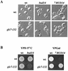
Bud14p and Glc7p share a function in polarized growth. (A) Morphology of double mutants. For the four left panels, wild-type (wt) (KT1357), bud14 (SY3899), glc7-132 (KT1706), and bud14 glc7-132 (SY3901) cells were grown to mid-log phase in YPD medium at 30°C and photographed at 100×. Arrows denote wide bud necks. For the two right panels, GAL-BUD14 (SY3899) and GAL-BUD14 glc7-132 (SY3901) cells were grown to mid-log phase in YPGal medium. Bar, 5 μm. (B) Glc7p and Bud14p share a function in cell growth. Equal concentrations of cells described for panel A were spotted onto the indicated media and incubated for 2 days at 30°C or at 37°C as indicated.
FIG. 7.
FIG. 7.
Glc7p is required for filamentous growth. (A) Plate-washing assay. Equal concentrations of wild-type (wt) (PAY704-1), glc7-10 (PAY700-4), glc7-12 (PAY701-3), and glc7-13 mutant (PAY702-4) cells were spotted onto YPD solid agar medium for 10 days at 30°C. The plate was photographed (left panel), washed, and photographed again (right panel). (B) Invaded cells were observed by microscopic examination of microcolonies on the washed YPD plate shown in panel A, and representative microcolonies were photographed. Bar, 20 μm.
FIG. 8.
FIG. 8.
Genetic interactions between Bud14p, Ste20p, Glc7p, and the polarisome (Bni1p), and their known roles in polarized growth, mating, and filamentous growth. Arrows denote positive functions, barred lines denote inhibitory functions, and the three stacked lines denote both genetic and physical interactions.

Similar articles

Cited by

References

    1. Ahn, S. H., A. Acurio, and S. J. Kron. 1999. Regulation of G2/M progression by the STE mitogen-activated protein kinase pathway in budding yeast filamentous growth. Mol. Biol. Cell 10:3301-3316. - PMC - PubMed
    1. Amberg, D. C., J. E. Zahner, J. W. Mulholland, J. R. Pringle, and D. Botstein. 1997. Aip3p/Bud6p, a yeast actin-interacting protein that is involved in morphogenesis and the selection of bipolar budding sites. Mol. Biol. Cell 8:729-753. - PMC - PubMed
    1. Andrews, P. D., and M. J. Stark. 2000. Type 1 protein phosphatase is required for maintenance of cell wall integrity, morphogenesis and cell cycle progression in Saccharomyces cerevisiae. J. Cell Sci. 113:507-520. - PubMed
    1. Bailis, J. M., and G. S. Roeder. 2000. Pachytene exit controlled by reversal of Mek1-dependent phosphorylation. Cell 101:211-221. - PubMed
    1. Baker, S. H., D. L. Frederick, A. Bloecher, and K. Tatchell. 1997. Alanine-scanning mutagenesis of protein phosphatase type 1 in the yeast Saccharomyces cerevisiae. Genetics 145:615-626. - PMC - PubMed

Publication types

MeSH terms