. 2002 Dec;1(6):1021-31.
doi: 10.1128/EC.1.6.1021-1031.2002.

Rvs161p and sphingolipids are required for actin repolarization following salt stress

Affiliations

Rvs161p and sphingolipids are required for actin repolarization following salt stress

Axelle Balguerie et al. Eukaryot Cell. 2002 Dec.

Abstract

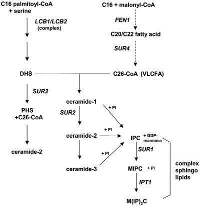
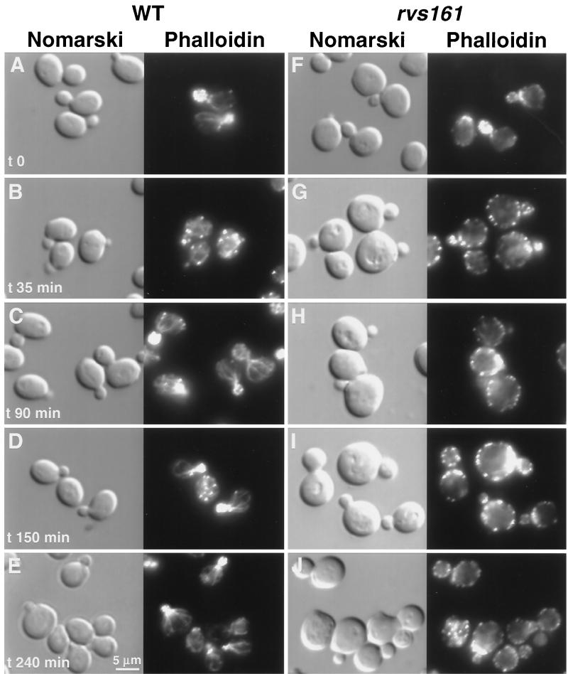
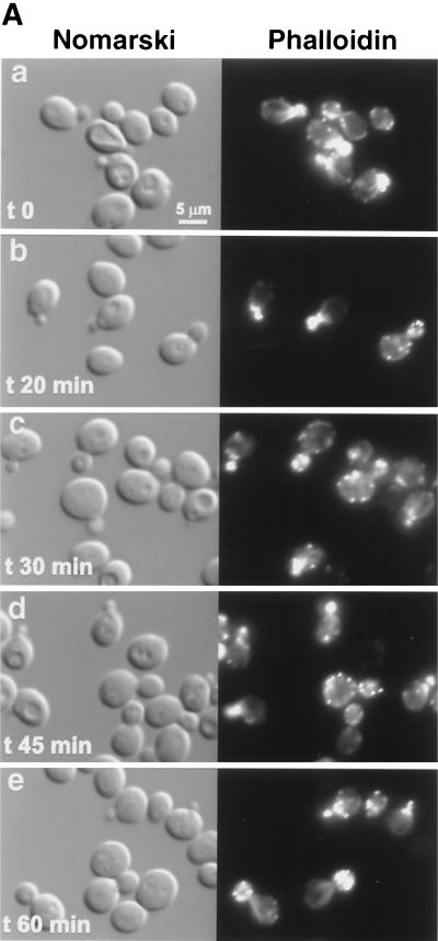
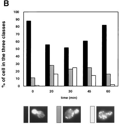
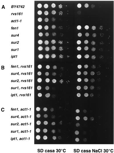
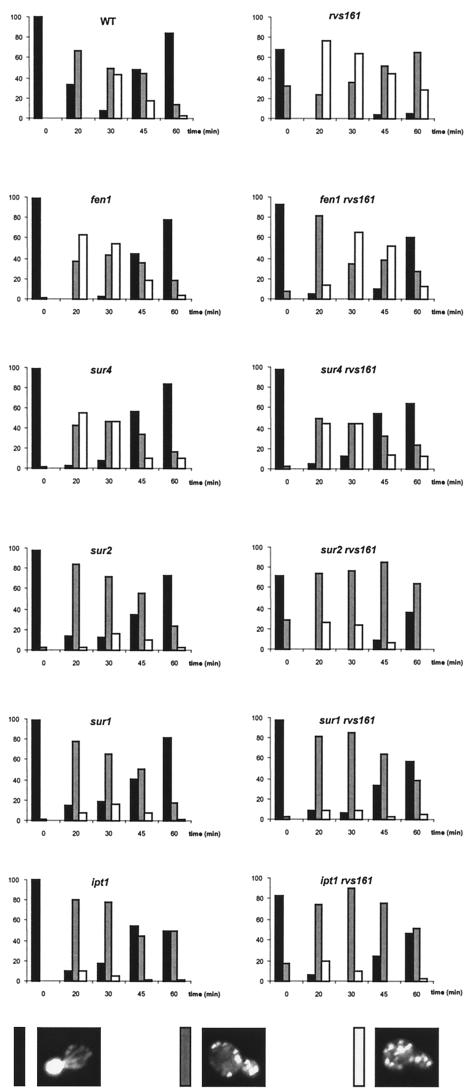
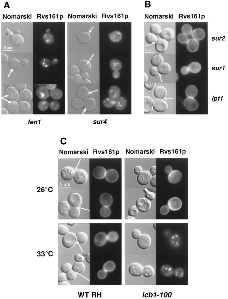
In Saccharomyces cerevisiae, the actin cytoskeleton is depolarized by NaCl stress. In this study, the response was maximal after 30 min, and then actin patches repolarized. Rvs161p was required for actin repolarization because the rvs161delta mutant did not repolarize actin patches after growth in a salt medium. Mutations suppressing the rvs161delta-related salt sensitivity all occurred in genes required for sphingolipid biosynthesis: FEN1, SUR4, SUR2, SUR1, and IPT1. These suppressors also suppressed act1-1-related salt sensitivity and the defect in actin repolarization of the rvs161delta mutant, providing a link between sphingolipids and actin polarization. Indeed, deletion of the suppressor genes suppressed the rvs161delta defect in actin repolarization in two ways: either actin was not depolarized at the wild-type level in a set of suppressor mutants, or actin was repolarized in the absence of Rvs161p in the other suppressor mutants. Rvs161p was localized as cortical patches that concentrated at polarization sites, i.e., bud emergence and septa, and was found to be associated with lipid rafts. An important link between sphingolipids and actin polarization is that Rvs161p was required for actin repolarization and was found to be located in lipid rafts.


Figures

FIG. 1.
FIG. 1.
Sphingolipid biosynthetic pathway in S. cerevisiae, adapted from Dickson and Lester (19) and Schneiter (37). Genes are shown in capital italic letters. LCB1 encodes a subunit of the serine palmitoyl transferase, FEN1 and SUR4 encode the elongases and are partially redundant, SUR2 encodes a hydroxylase, SUR1 encodes a mannosyl transferase, and IPT1 encodes an inositol phosphotransferase. Abbreviations: CoA, coenzyme A; VLCFA, very long chain fatty acid; PI, phosphatidylinositol; DHS and PHS, sphingoid bases dihydrosphingosine and phytosphingosine, respectively. IPC, inositolphosphorylceramide; MIPC, mannosyl-inositolphosphorylceramide; and M(IP)2C, mannosyl-diinositolphosphorylceramide.
FIG. 2.
FIG. 2.
Rvs161p is required for actin repolarization following salt stress. Wild-type (WT, A to E) and rvs161Δ mutant (F to J) strains were grown in YPD at 30°C, and at time zero, NaCl was added to a final concentration of 3.4%. Aliquots were removed at different times as indicated. Samples were fixed, stained with Alexa-phalloidin, and observed by fluorescence and Nomarski microscopy to visualize the actin cytoskeleton and whole cells, respectively.
FIG. 3.
FIG. 3.
wsc1 mutant impaired for actin cytoskeleton depolarization following salt stress. (A) The wsc1 strain was grown and treated as described for Fig. 2. (B) Cells (≈100) were classified and quantified according to their polarization state. Only cells with small buds were scored. Cells with actin patches concentrated in the small bud, with fewer than four patches in the mother cell, were classified as polarized cells (black bars). Cells with actin patches concentrated in the small bud but with more than four patches in the mother cell were classified as partially depolarized cells (grey bars). Cells with more actin patches in the mother cell than in the small bud were classified as totally depolarized cells (white bars).
FIG. 3.
FIG. 3.
wsc1 mutant impaired for actin cytoskeleton depolarization following salt stress. (A) The wsc1 strain was grown and treated as described for Fig. 2. (B) Cells (≈100) were classified and quantified according to their polarization state. Only cells with small buds were scored. Cells with actin patches concentrated in the small bud, with fewer than four patches in the mother cell, were classified as polarized cells (black bars). Cells with actin patches concentrated in the small bud but with more than four patches in the mother cell were classified as partially depolarized cells (grey bars). Cells with more actin patches in the mother cell than in the small bud were classified as totally depolarized cells (white bars).
FIG. 4.
FIG. 4.
fen1, sur4, sur2, sur1, and ipt1 mutants suppress the salt sensitivity of the rvs161Δ and act1-1 mutants. The growth phenotypes were evaluated following a serial dilution drop test. The undiluted drop contained 5 × 10⁴ cells. The complete genotype of the strains is given in Table 1.
FIG. 5.
FIG. 5.
Sphingolipid mutants are impaired in actin depolarization and suppress the repolarization defect of the rvs161Δ mutant. The wild-type (WT) and the indicated mutant strains were grown as described for Fig. 2. Cells (≈100) were classified and quantified according to their polarization state as in Fig. 3.
FIG. 6.
FIG. 6.
Rvs161p-GFP localizes as cortical patches and concentrates at polarization sites. Wild-type cells grown in SD-casa medium were observed by Nomarski and fluorescence microscopy to visualize whole cells and Rvs161p-GFP localization, respectively. The equatorial view represents the middle of the cell. For the top view, the focusing was done on the top of the cell. (A) Unbudded cell, (B) bud emergence cell, and (C) budded cell with the septum. The arrow on the Nomarski image indicates the septum between the mother cell and the bud.
FIG. 7.
FIG. 7.
Rvs161p-GFP localization is impaired in fen1, sur4, and lcb1-100 mutants. Different mutant cells grown in SD-casa medium were observed by Nomarski and fluorescence microscopy to visualize whole cells and Rvs161p-GFP localization, respectively. (A) In the fen1 and sur4 mutants, the arrow on the Nomarski images indicates the structures that contain Rvs161p-GFP. (B) In the sur2, sur1, and ipt1 mutants, the arrow on the Nomarski images indicates the septum between the mother cell and the bud. (C) Rvs161p-GFP localization in the lcb1-100 mutant and the corresponding wild-type (RH1800) cells grown at 26°C and shifted to 33°C for 90 min. The arrow on the Nomarski images indicates the septum between the mother cell and the bud.
FIG. 8.
FIG. 8.
Pma1p-GFP localization is affected in the fen1 and sur4 mutants. The indicated strains were grown in SD-casa medium and observed by Nomarski and fluorescence microscopy to visualize whole cells and Pma1p-GFP localization, respectively. WT, wild type.
FIG. 9.
FIG. 9.
Rvs161p is associated with DRMs. (A) Wild-type cells were treated as described in Materials and Methods. Rvs161p was immunoprecipitated from the membrane (M), the detergent-resistant membrane (R), and the respective soluble (S) fractions with specific antibodies, followed by SDS-PAGE and Phosphorimager analysis. (B) The lcb1-100 mutant and the corresponding wild-type (WT, RH1800) strain were grown at the permissive (24°C) or the restrictive (37°C) temperature and treated as described in Materials and Methods. Rvs161p was immunoprecipitated from detergent-resistant membrane (R) and soluble (S) fractions with specific antibodies and analyzed by SDS-PAGE and Phosphorimager analysis.

References

    1. Bagnat, M., S. Keränen, A. Shevchenko, A. Shevchenko, and K. Simons. 2000. Lipid rafts function in biosynthetic delivery of proteins to the cell surface in yeast. Proc. Natl. Acad. Sci. USA 97:3254-3259. - PMC - PubMed
    1. Bagnat, M., M. Chang, and K. Simons. 2001. Plasma membrane proton ATPase Pma1p requires raft association for surface delivery in yeast. Mol. Biol. Cell 12:4129-4138. - PMC - PubMed
    1. Bauer, F., M. Urdaci, M. Aigle, and M. Crouzet. 1993. Alteration of a yeast SH3 protein leads to conditional viability with defects in cytoskeletal and budding patterns. Mol. Cell. Biol. 13:5070-5084. - PMC - PubMed
    1. Bonneaud, N., O. Ozier-Kalogeropoulos, G. Y. Li, M. Labouesse, L. Minvielle-Sebastia, and F. Lacroute. 1991. A family of low and high copy replicative, integrative and single-stranded S. cerevisiae/E. coli shuttle vectors. Yeast 7:609-615. - PubMed
    1. Breton, A. M., and M. Aigle. 1998. Genetic and functional relationship between Rvsp, myosin and actin in Saccharomyces cerevisiae. Curr. Genet. 34:280-286. - PubMed

Publication types

MeSH terms