. 2003 Jan;77(1):45-56.
doi: 10.1128/jvi.77.1.45-56.2003.

The major apoptotic pathway activated and suppressed by poliovirus

Affiliations

The major apoptotic pathway activated and suppressed by poliovirus

George A Belov et al. J Virol. 2003 Jan.

Abstract

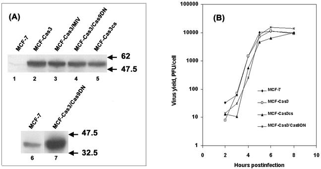
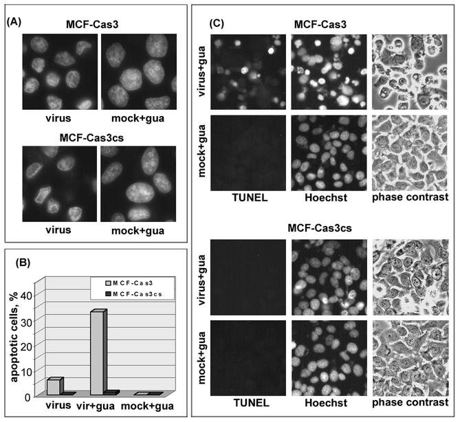
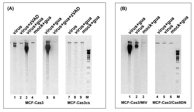
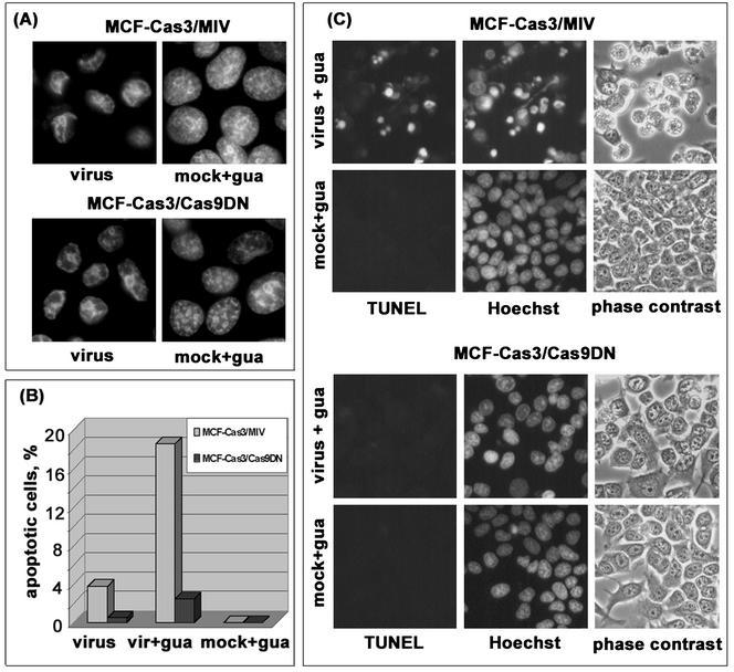
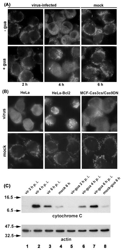
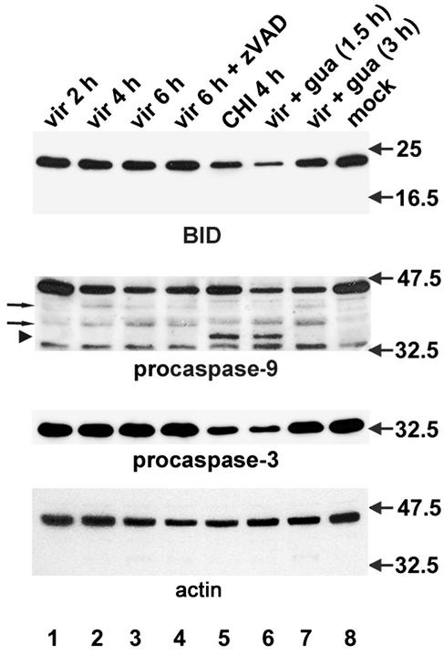
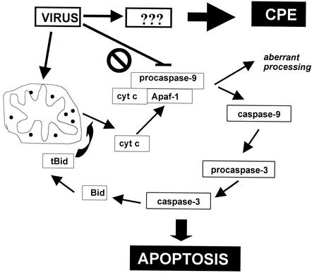
Cells respond to poliovirus infection by switching on the apoptotic program, implementation of which is usually suppressed by viral antiapoptotic functions. We show here that poliovirus infection of HeLa cells or derivatives of MCF-7 cells was accompanied by the efflux of cytochrome c from mitochondria. This efflux occurred during both abortive infection (e.g., interrupted by guanidine-HCl and ending with apoptosis) and productive infection (leading to cytopathic effect). The former type of infection, but not the latter, was accompanied by truncation of the proapoptotic protein Bid. The virus-triggered cytochrome c efflux was suppressed by overexpression of Bcl-2. Both abortive and productive infections also resulted in a decreased level of procaspase-9, as revealed by Western blotting. In the former case, this decrease was accompanied by the accumulation of a protein with the electrophoretic mobility of active caspase-9. In contrast, in the productively infected cells, the latter protein was absent but caspase-9-related polypeptides with altered mobility could be detected. Both caspase-9 and caspase-3 were shown to be essential for the development of such hallmarks of virus-induced apoptosis as chromatin condensation, DNA degradation, and nuclear fragmentation. These and some other results suggest the following scenario. Poliovirus infection activates the apoptotic pathway, involving mitochondrial damage, cytochrome c efflux, and consecutive activation of caspase-9 and caspase-3. The apoptotic signal appears to be amplified by a loop which includes secondary processing of Bid. The implementation of the apoptotic program in productively infected cells may be suppressed, however, by the viral antiapoptotic functions, which act at a step(s) downstream of the cytochrome c efflux. The suppression appears to be caused, at least in part, by aberrant processing and degradation of procaspase-9.


Figures

FIG. 1.
FIG. 1.
General properties of MCF-7 cell derivatives. (A) Overexpression of relevant caspase species in the derived cell lines as revealed by Western blotting. Total cell lysates prepared from the respective cells were probed with polyclonal antibodies against procaspase-3 (lanes 1 to 5) and procaspase-9 (lanes 6 and 7). The caspase-3-specific bands, seen in the MCF-7 derivatives, correspond to the proenzyme fused to GFP. (B) Single-cycle growth curves of poliovirus in MCF-7, MCF-Cas3, MCF-Cas3cs, and MCF-Cas3/Cas9DN cells. The infection was carried out as described in Materials and Methods. The harvest was plaque titrated on RD cells.
FIG. 2.
FIG. 2.
Responses of MCF-Cas3 and MCF-Cas3cs cells to productive and abortive poliovirus infection. (A) Nuclear CPE in productively infected cells. Hoechst 33342 was added at 5.5 h p.i., and the cells were fixed at 6 h p.i. mock+gua, mock-infected cells with 100 μg of guanidine-HCl/ml added. (B) Percentages of TUNEL-positive infected cells at 6 h p.i. in the absence (virus) and presence (vir+gua) of 100 μg of guanidine-HCl/ml added at 2 h p.i. The data represent the averages of two experiments. (C) Development of apoptosis in MCF-Cas3 but not in MCF-Cas3cs cells upon abortive infection. Guanidine-HCl (100 μg/ml) was added to the virus-infected (virus+gua) and mock-infected (mock+gua) cells at 2 h p.i. Hoechst 33342 was added at 5.5 h p.i. The cells were fixed at 6 h p.i. and processed for the TUNEL assay. Qualitatively similar results were obtained when productive infection was interrupted by guanidine-HCl at 1.5 h p.i. (not shown).
FIG. 3.
FIG. 3.
Electrophoretic assay for DNA degradation upon productive (virus) and abortive (virus+gua) poliovirus infection of MCF-Cas3, MCF-Cas3cs, MCF-Cas3/MIV, and MCF-Cas-3/Cas9DN cells. Guanidine-HCl was added, when appropriate, 2 h after infection (virus+gua) or mock infection (mock+gua). To the sample analyzed in lanes 2 and 6, 100 μM zVAD.fmk (zVAD) was added at the onset of infection. The cells were harvested at 6 h p.i., except for the samples in lanes 5 and 6, where the cells were harvested at 8 h p.i. Lane M shows a PstI digest of λ phage DNA.
FIG. 4.
FIG. 4.
Responses of MCF-Cas3/MIV and MCF-Cas3/Cas9DN cells to productive and abortive poliovirus infections. (A) Nuclear CPE in productively infected cells. Hoechst 33342 was added at 7.5 h p.i., and the cells were fixed at 8 h p.i. mock+gua, mock-infected cells with 100 μg of guanidine-HCl/ml added. (B) Percentages of TUNEL-positive infected cells at 8 h p.i. in the absence (virus) and presence (vir+gua) of 100 μg of guanidine-HCl/ml added at 2 h p.i. The data represent the averages of two experiments. (C) Development of apoptosis in MCF-Cas3/MIV but not MCF-Cas3/Cas9DN cells. Guanidine-HCl (100 μg/ml) was added to the virus-infected (virus + gua) and mock-infected (mock+gua) cells at 2 h p.i. Hoechst 33342 was added at 7.5 h p.i. The cells were fixed at 8 h p.i. and processed for the TUNEL assay. Qualitatively similar results were obtained when productive infection was interrupted by guanidine-HCl at 1.5 h p.i. (not shown).
FIG. 5.
FIG. 5.
Cytochrome c efflux upon productive and abortive poliovirus infection. (A) Virus-infected and mock-infected HeLa cells were incubated for different time intervals in the absence (− gua) and presence (+ gua) of 100 μg of guanidine-HCl/ml (added at 1.5 h p.i.). (B) Productively infected and mock-infected HeLa, HeLa-Bcl2, and MCF-Cas3/Cas9DN cells were incubated for 6 h. Immunostaining for cytochrome c was performed as described in Materials and Methods. (C) Western blot analysis of the soluble cytoplasmic fraction (see Materials and Methods) from HeLa cells productively (vir) and abortively (vir-gua) infected as for panel A. A decrease in the content of cytochrome c in the 6-h sample (lane 3) compared to the 4-h sample (lane 2) could be explained by the lysis of a proportion of cells due to CPE caused by productive infection.
FIG. 6.
FIG. 6.
Processing of Bid, procaspase-9, and procaspase-3 in virus-infected and uninfected HeLa cells. Productively infected cells (vir) were incubated for 2, 4, and 6 h p.i. (lanes 1 to 3). Infection in the sample in lane 4 was carried out in the presence of 100 μM zVAD.fmk (+ zVAD). In the samples in lanes 6 and 7, productive infection was interrupted by the addition of 100 μg of guanidine-HCl/ml (+ gua) at 1.5 and 3 h p.i., respectively, and the cells were harvested at 6 h p.i. In the sample in lane 5, uninfected cells were incubated in the presence of 100 μg of CHI/ml for 4 h. The sample in lane 8 represents mock-infected cells. Western blot analysis of a cellular extract was performed as described in Materials and Methods. The positions of normally and aberrantly processed caspase-9 are marked by the arrowhead and arrows, respectively. The positions of protein markers are also indicated.
FIG. 7.
FIG. 7.
Schematic representation of the major poliovirus-triggered apoptotic pathway as characterized in the present study. Poliovirus infection results in mitochondrial damage and efflux of cytochrome c (cyt c) into the cytoplasm. Together with Apaf-1 and cytochrome c, procaspase-9 forms an apoptosome in a dATP-dependent reaction, followed by generation of active caspase-9. The latter cleaves procaspase-3, with generation of active caspase-3. This effector protease destroys various important substrates and activates other hydrolases, e.g., DNases, and eventually leads to apoptotic death. The apoptotic signal appears to be amplified through limited proteolysis of Bid by caspase-3. The truncated form of Bid (tBid), in cooperation with some proapoptotic factors, causes further mitochondrial damage. Concurrently, a viral antiapoptotic function promotes aberrant processing of procaspase-9, hindering its activation and resulting in the development of CPE. The step at which this aberrant processing occurs has yet to be defined.

References

    1. Adams, J. M., and S. Cory. 2001. Life-or-death decisions by the Bcl-2 protein family. Trends Biochem. Sci. 26:61-66. - PubMed
    1. Adrain, C., and S. J. Martin. 2001. The mitochondrial apoptosome: a killer unleashed by the cytochrome seas. Trends Biochem. Sci. 26:390-397. - PubMed
    1. Agol, V. I., G. A. Belov, K. Bienz, D. Egger, M. S. Kolesnikova, N. T. Raikhlin, L. I. Romanova, E. A. Smirnova, and E. A. Tolskaya. 1998. Two types of death of poliovirus-infected cells: caspase involvement in the apoptosis but not cytopathic effect. Virology 252:342-353. - PubMed
    1. Agol, V. I., G. A. Belov, K. Bienz, D. Egger, M. S. Kolesnikova, L. I. Romanova, L. V. Sladkova, and E. A. Tolskaya. 2000. Competing death programs in poliovirus-infected cells: commitment switch in the middle of the infectious cycle. J. Virol. 74:5534-5541. - PMC - PubMed
    1. Ammendolia, M. G., A. Tinari, A. Calcabrini, and F. Superti. 1999. Poliovirus infection induces apoptosis in CaCo-2 cells. J. Med. Virol. 59:122-129. - PubMed

Publication types