. 2003 Jan;77(1):217-27.
doi: 10.1128/jvi.77.1.217-227.2003.

Novel member of the CD209 (DC-SIGN) gene family in primates

Affiliations

Novel member of the CD209 (DC-SIGN) gene family in primates

Arman A Bashirova et al. J Virol. 2003 Jan.

Abstract

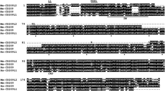
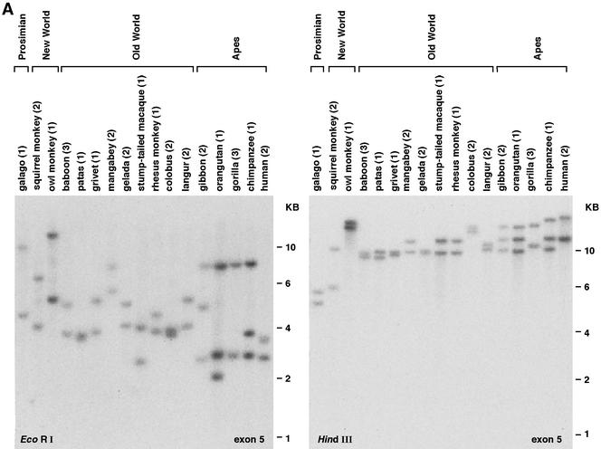
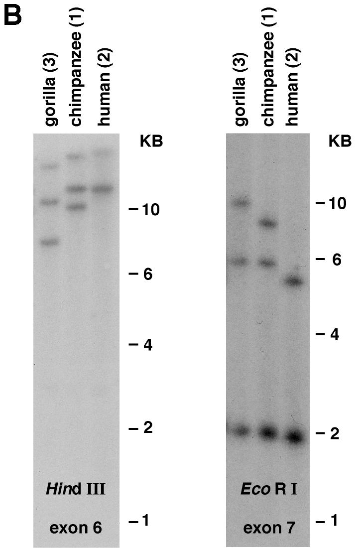
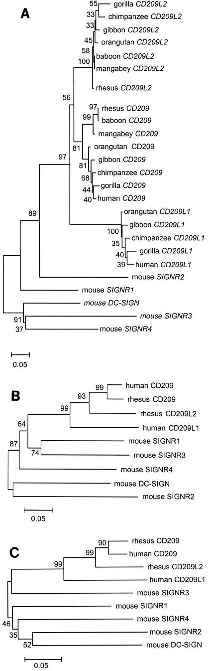
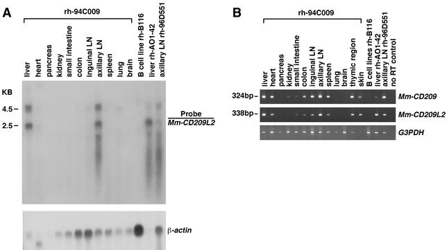
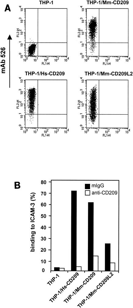
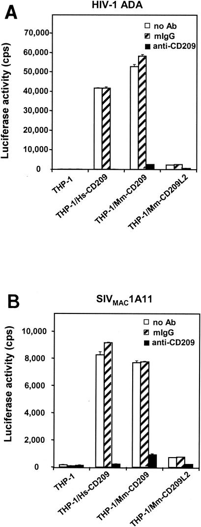
Two CD209 family genes identified in humans, CD209 (DC-SIGN) and CD209L (DC-SIGNR/L-SIGN), encode C-type lectins that serve as adhesion receptors for ICAM-2 and ICAM-3 and participate in the transmission of human and simian immunodeficiency viruses (HIV and SIV, respectively) to target cells in vitro. Here we characterize the CD209 gene family in nonhuman primates and show that recent evolutionary alterations have occurred in this family across primate species. All of the primate species tested, specifically, Old World monkeys (OWM) and apes, have orthologues of human CD209. In contrast, CD209L is missing in OWM but present in apes. A third family member, that we have named CD209L2, was cloned from rhesus monkey cDNA and subsequently identified in OWM and apes but not in humans. Rhesus CD209L2 mRNA was prominently expressed in the liver and axillary lymph nodes, although preliminary data suggest that levels of expression may vary among individuals. Despite a high level of sequence similarity to both human and rhesus CD209, rhesus CD209L2 was substantially less effective at binding ICAM-3 and poorly transmitted HIV type 1 and SIV to target cells relative to CD209. Our data suggest that the CD209 gene family has undergone recent evolutionary processes involving duplications and deletions, the latter of which may be tolerated because of potentially redundant functional activities of the molecules encoded by these genes.


Figures

FIG. 1.
FIG. 1.
Amino acid alignment of putative Mm-CD209, Mm-CD209L2, human CD209 (Hs-CD209), and Hs-CD209L1. The potential internalization motifs, LL and YXXL, are annotated in the cytoplasmic domain, and the transmembrane domain (TM) is underlined. The starts of the repeats in the neck region (*) and the beginning of the CRD are indicated below the sequences.
FIG. 2.
FIG. 2.
Southern blot analysis of primate genomic DNA. Hybridization patterns suggest the presence of two or three CD209 family genes in various primate species. The number of animals of each genus tested is indicated in parentheses. Individuals from the same genus either showed the same banding pattern (most cases) or revealed restriction fragment length polymorphisms (see supplemental Fig. 1 at URL address http:/home.ncifcrf.gov/ccr/lgd/cd209/sup_data.htm). (A) Hybridization with the human CD209 exon 5 probe. (B) Hybridization with the exon 6 and exon 7 probes showing three bands in the gorilla sample.
FIG. 2.
FIG. 2.
Southern blot analysis of primate genomic DNA. Hybridization patterns suggest the presence of two or three CD209 family genes in various primate species. The number of animals of each genus tested is indicated in parentheses. Individuals from the same genus either showed the same banding pattern (most cases) or revealed restriction fragment length polymorphisms (see supplemental Fig. 1 at URL address http:/home.ncifcrf.gov/ccr/lgd/cd209/sup_data.htm). (A) Hybridization with the human CD209 exon 5 probe. (B) Hybridization with the exon 6 and exon 7 probes showing three bands in the gorilla sample.
FIG. 3.
FIG. 3.
Phylogenetic analysis of the CD209 gene family. (A) Neighbor-joining tree of nucleotide sequences homologous to a 249-bp fragment of human CD209 exon 7. Two to four individuals from each genus were sequenced. (B) Neighbor-joining tree of putative full-length amino acid sequences of the CD209 family proteins. (C) Neighbor-joining tree of amino acid sequences of the CRDs in the CD209 family proteins.
FIG. 4.
FIG. 4.
Expression of the Mm-CD209L2 gene in rhesus monkey tissues. (A) Northern analysis of the Mm-CD209L2 gene. Total RNAs isolated from various tissues and a B-cell line were hybridized with a 794-bp Mm-CD209L2 cDNA probe. The 2.5-kb band is presumably Mm-CD209L2 mRNA, and the 4.5-kb doublet most likely corresponds to Mm-CD209. A human β-actin probe was used as a control for RNA loading. (B) RT-PCR analysis of Mm-CD209 and Mm-CD209L2 expression. Gene-specific primers were used to analyze expression of the two genes. In addition to the RNA samples represented in panel A, skin and thymic region RNAs were also included. Human G3PDH primers were used as a positive control. Four individual rhesus monkeys (rh-94C009, rh-A01-42, rh-95D551, and rh-B116) are represented.
FIG. 5.
FIG. 5.
Cell lines expressing CD209 family molecules bind ICAM-3. (A) THP-1 monocytes stably transduced with human and macaque CD209 family molecules were assessed for protein expression with cross-reactive MAb 526. Positive expression is indicated on the FL-2 axis. Isotypic antibody control stainings of the different cell lines were uniformly negative. (B) Adhesion of ICAM-3 to THP-1/Hs-CD209, THP-1/Hs-CD209L, THP-1/Mm-CD209, and THP-1/Mm-CD209L2 cells was measured by FACS analysis with a fluorescent bead adhesion assay as previously described (27). Control mouse IgG or CD209 MAb 526 (10 μg/ml) was preincubated with cells. Adhesion of ICAM-3 to THP-1 parental cells was less than 5%. The results of one experiment representative of two are shown.
FIG. 6.
FIG. 6.
Impaired virus transmission by Mm-CD209L2. Relative virus capture and transmission by CD209 family molecules was assayed with HIV-Luc pseudotyped with HIV-1 ADA Env (A) or SIVMAC1A11 Env (B). THP-1, THP-1/Hs-CD209, THP-1/Mm-CD209, and THP-1/Mm-CD209L2 donor cells were preincubated with the MAbs (10 μg/ml) for 30 min at 37°C, after which HIV-Luc pseudotypes were added and the mixture was incubated for an additional 3 h at 37°C. The cells were then washed and cocultured with Hut/CCR5 target cells in the presence of Polybrene. HIV-1 infection was determined after 2 days by measuring the luciferase activity. Treatment with MAb 526 was used to evaluate the necessity of CD209 molecules for transmission, and mouse IgG (mIgG) was used as a nonspecific antibody control. Each set of data represents the mean of three separate wells of infected cells. The results of one experiment representative of three are shown. cps, counts per second.

References

    1. Baribaud, F., S. Pohlmann, T. Sparwasser, M. T. Kimata, Y. K. Choi, B. S. Haggarty, N. Ahmad, T. Macfarlan, T. G. Edwards, G. J. Leslie, J. Arnason, T. A. Reinhart, J. T. Kimata, D. R. Littman, J. A. Hoxie, and R. W. Doms. 2001. Functional and antigenic characterization of human, rhesus macaque, pigtailed macaque, and murine DC-SIGN. J. Virol. 75:10281-10289. - PMC - PubMed
    1. Bashirova, A. A., T. B. Geijtenbeek, G. C. van Duijnhoven, S. J. van Vliet, J. B. Eilering, M. P. Martin, L. Wu, T. D. Martin, N. Viebig, P. A. Knolle, V. N. KewalRamani, Y. van Kooyk, and M. Carrington. 2001. A dendritic cell-specific intercellular adhesion molecule 3-grabbing nonintegrin (DC-SIGN)-related protein is highly expressed on human liver sinusoidal endothelial cells and promotes HIV-1 infection. J. Exp. Med. 193:671-678. - PMC - PubMed
    1. Benveniste, R. E., R. Heinemann, G. L. Wilson, R. Callahan, and G. J. Todaro. 1974. Detection of baboon type C viral sequences in various primate tissues by molecular hybridization. J. Virol. 14:56-67. - PMC - PubMed
    1. Benveniste, R. E., L. Kuller, S. T. Roodman, S. L. Hu, and W. R. Morton. 1993. Long-term protection of macaques against high-dose type D retrovirus challenge after immunization with recombinant vaccinia virus expressing envelope glycoproteins. J. Med. Primatol. 22:74-79. - PubMed
    1. Blauvelt, A., S. Glushakova, and L. B. Margolis. 2000. HIV-infected human Langerhans cells transmit infection to human lymphoid tissue ex vivo. AIDS 14:647-651. - PubMed

Publication types

MeSH terms

Substances

LinkOut - more resources