. 2003 Jan;77(1):382-90.
doi: 10.1128/jvi.77.1.382-390.2003.

DNA immunization with hepatitis C virus (HCV) polycistronic genes or immunization by HCV DNA priming-recombinant canarypox virus boosting induces immune responses and protection from recombinant HCV-vaccinia virus infection in HLA-A2.1-transgenic mice

Affiliations

DNA immunization with hepatitis C virus (HCV) polycistronic genes or immunization by HCV DNA priming-recombinant canarypox virus boosting induces immune responses and protection from recombinant HCV-vaccinia virus infection in HLA-A2.1-transgenic mice

Preeti Pancholi et al. J Virol. 2003 Jan.

Abstract

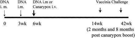
We studied immune responses to hepatitis C virus (HCV) genes delivered as DNA encoding the entire HCV protein coding genome in two polycistronic plasmids encoding HCV capsid-E1-E2-NS2-NS3 and HCV NS3-NS4-NS5 in HLA-A2.1-transgenic mice. Immune responses to HCV DNA prime and recombinant canarypox virus boost were also studied with the above constructs. At 8 weeks after a canarypox virus boost, the DNA prime/canarypox virus boosting regimen induced potent cellular immune responses to HCV structural and nonstructural proteins on target cells expressing the HLA-A2.1 allele. High frequencies of gamma interferon-secreting cells, as detected by enzyme-linked immunospot assay, were obtained in response to several endogenously expressed HCV proteins. We also observed cytotoxic-T-lymphocyte reactivity in response to endogenously expressed HCV proteins in fresh spleen cells without in vitro expansion. Upon challenge with a recombinant vaccinia virus expressing HCV proteins at 2 months postimmunization, the HCV DNA prime/canarypox virus-immunized mice showed a complete reduction in vaccinia virus titers compared to HCV DNA prime/boost- and mock-immunized controls. Immune responses were still detectable 4 months after canarypox virus boost in immunized mice. Interestingly, at 10 months postimmunization (8 months after canarypox virus boost), the protection in HCV DNA prime/boost-immunized mice against recombinant HCV-vaccinia virus challenge was higher than that observed in HCV DNA prime/canarypox virus boost-immunized mice.


Figures

FIG. 1.
FIG. 1.
HCV DNA/canarypox virus immunization and challenge scheme. HLA-A2.1 mice were immunized with HCV plasmids HCV (C-NS3) and HCV (NS3-NS5) at time zero and at 3 weeks. The animals were divided into two groups. At 3 weeks after the second DNA immunization, half of the animals were boosted with HCV DNA plasmids and the other half received recombinant HCV canarypox expressing the same HCV proteins. At 2 and 8 months after the canarypox virus boost, the HCV DNA prime/boost, and the HCV DNA prime/canarypox virus boost mice were challenged with 107 PFU each of vaccinia virus encoding HCV C-NS3 and vaccinia virus encoding HCV NS3-NS5 i.p. to determine vaccinia virus titers in mouse ovaries.
FIG. 2.
FIG. 2.
Cytokine gene expression. Mouse splenocytes were analyzed for TCR αβ, CD4, and CD8 and for IL-2, IFN-γ, and TNF-α cytokine mRNA gene expression at 8 weeks after canarypox virus boost by the RNase protection assay. Splenocytes were stimulated with stimulator cells infected with recombinant vaccinia virus parent or recombinant vaccinia virus expressing HCV C-NS3 or recombinant vaccinia virus expressing β-Gal proteins. Mouse GAPDH and L32 genes were used for normalization. Mice immunized with HCV DNA prime/boost (lane 3) and HCV DNA prime/canarypox virus boost (lane 7) showed increased mRNA levels of IFN-γ gene expression after in vitro stimulation with target cells expressing HCV proteins. The increase in the levels of IFN-γ was specific to HCV and was not obtained with target cells expressing β-Gal protein (lanes 1 and 5) or target cells infected with recombinant vaccinia virus parent (lanes 2 and 6). Lanes 4 and 8 represent unstimulated splenocytes from HCV DNA prime/boost and HCV DNA prime/canarypox virus boost mice, respectively.
FIG. 3.
FIG. 3.
Direct ex vivo CTL responses in HLA-A2.1 mice immunized with plasmid DNA construct encoding HCV C-NS3 and HCV NS3-NS5: effect of canarypox virus boost. HLA-A2.1 mice were immunized with HCV plasmid pRC/C-NS3 and pRC/NS3-NS5 DNA. Half of the animals were boosted with canarypox virus. CTL responses with unstimulated spleen cells were analyzed 8 weeks after canarypox virus boost with EA2Kb target cells infected with vaccinia virus-encoded HCV proteins. Panel A (HCV DNA prime and boost) shows specific CTL lysis by effector cells at different E:T ratios from HLA-A2.1 mice after HCV DNA prime and boost against EA2Kb target cells infected with the vaccinia virus (WR)-encoded HCV gene products capsid, envelope E1, envelope E2, NS2, and NS3. Panel B (HCV DNA prime/canarypox virus boost) likewise shows the specific lysis of target cells after HCV DNA prime/canarypox virus boost immunization.
FIG. 4.
FIG. 4.
In vitro-expanded CTL responses in HLA-A2.1 mice immunized with HCV plasmid DNA encoding C-NS3 and NS3-NS5 proteins: effect of canarypox virus boost. HLA-A2.1 mice were immunized with HCV plasmid pRC/C-NS3 and pRC/NS3-NS5 DNA. Half of the animals were boosted with canarypox virus. CTL responses were analyzed 8 weeks after a canarypox virus boost with in vitro-stimulated spleen cells with EA2Kb target cells infected with vaccinia virus-encoded HCV proteins. Panel A (HCV DNA prime and boost) shows specific CTL lysis by effector cells at different E:T ratios from HLA-A2.1 mice after HCV DNA prime and boost against EA2Kb target cells infected with the vaccinia virus (WR)-encoded HCV gene products capsid, envelope E1, envelope E2, NS2, and NS3. Panel B (HCV DNA prime/canarypox virus boost) likewise shows the specific lysis of target cells after HCV DNA prime/canarypox virus boost immunization.
FIG. 5.
FIG. 5.
Blocking of CTL responses in HLA-A2.1 mice against HCV-expressing targets by anti-CD8, anti-CD4, or anti-HLA-A2.1 antibodies. Mice were immunized with either control (empty plasmid) vector or HCV plasmid encoding pRC/C-NS3 + pRC/NS3-NS5. Half of the animals in each group were boosted with canarypox virus expressing the same HCV proteins. CTL responses were analyzed 8 weeks after canarypox virus boost after in vitro expansion for 5 days (expanded cells) by using EA2Kb target cells infected with vaccinia virus-encoded HCV C-NS3 proteins (WR C-NS3). As a control, lysis was also assessed against vaccinia virus parent [WR (P)]-infected target cells and against vaccinia virus expressing β-Gal protein. All responses shown are at an E:T ratio of 50:1. The blocking antibodies were present throughout the 5-h 51Cr release assay. The background lysis of target cells was ≤5%.
FIG. 6.
FIG. 6.
Vaccinia virus titers in ovaries 8 weeks after canarypox virus boost. At 8 weeks after a canarypox virus boost, HLA-A2.1 mice immunized with HCV DNA prime/boost or with HCV DNA prime/canarypox virus boost were challenged with 107 PFU each of vaccinia virus encoding HCV C-NS3 and vaccinia virus encoding HCV NS3-NS5 i.p. As controls, mice were challenged with recombinant vaccinia virus expressing β-Gal protein. The ovaries were harvested 5 days after challenge, and vaccinia virus titers were determined by plaque assay on BSC-40 cells.
FIG. 7.
FIG. 7.
Vaccinia virus titers in ovaries 8 months after a canarypox virus boost. At 8 months after a canarypox virus boost, HLA-A2.1 mice immunized with HCV DNA prime/boost or with HCV DNA prime/canarypox virus boost were challenged with 107 PFU each of vaccinia virus encoding HCV C-NS3 and vaccinia virus encoding HCV NS3-NS5 i.p. As controls, mice were challenged with recombinant vaccinia virus expressing β-Gal protein. The ovaries were harvested 5 days after challenge, and vaccinia virus titers were determined by plaque assay on BSC-40 cells.

References

    1. Alexander-Miller, M. A., G. R Leggatt, and J. A. Berzofsky. 1996. Selective expansion of high- or low-avidity cytotoxic T lymphocytes and efficacy for adoptive immunotherapy. Proc. Natl. Acad. Sci. USA 93:4102-4107. - PMC - PubMed
    1. Allen, T. M., T. U Vogel, D. H. Fuller, B. R. Mothe, S. Steffen, J. E. Boyson, T. Shipley, J. Fuller, T. Hanke, A. Sette, J. D. Altman, B. Moss, A. J. McMichael, and D. I. Watkins. 2000. Induction of AIDS virus-specific CTL activity in fresh, unstimulated peripheral blood lymphocytes from rhesus macaques vaccinated with a DNA prime/modified vaccinia virus Ankara boost regimen. J. Immunol. 164:4968-4978. - PubMed
    1. Alter, M. J., H. S. Margolis, K. Krawczynski, et al. 1992. The natural history of community-acquired hepatitis in the United States: the sentinel counties chronic non-A, non-B hepatitis study team. N. Engl. J. Med. 327:1899-1905. - PubMed
    1. Arichi, T., T. Saito, M. E. Major, I. M. Belyakov, M. Shirai, V. H. Engelhard, S. M. Feinstone, and J. A. Berzofsky. 2000. Prophylactic DNA vaccine for hepatitis C virus (HCV) infection: HCV-specific cytotoxic T lymphocyte induction and protection from HCV-recombinant vaccinia infection in an HLA-A2.1 transgenic mouse model. Proc. Natl. Acad. Sci. USA 97:297-302. - PMC - PubMed
    1. Bukh, J., R. H. Miller, and R. H. Purcell. 1995. Genetic heterogeneity of hepatitis C virus: quasispecies and genotypes. Semin. Liver Dis. 15:41-63. - PubMed

Publication types

MeSH terms