. 2003 Jan;77(1):673-84.
doi: 10.1128/jvi.77.1.673-684.2003.

The ND10 component promyelocytic leukemia protein relocates to human papillomavirus type 1 E4 intranuclear inclusion bodies in cultured keratinocytes and in warts

Affiliations

The ND10 component promyelocytic leukemia protein relocates to human papillomavirus type 1 E4 intranuclear inclusion bodies in cultured keratinocytes and in warts

Sally Roberts et al. J Virol. 2003 Jan.

Abstract

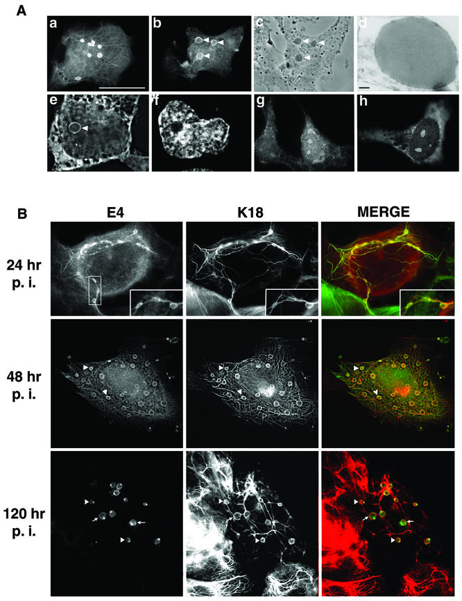
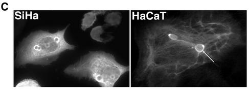
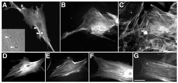
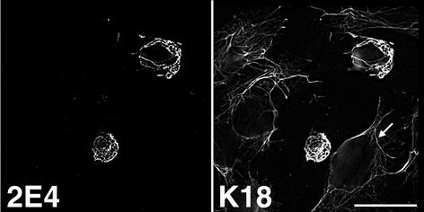
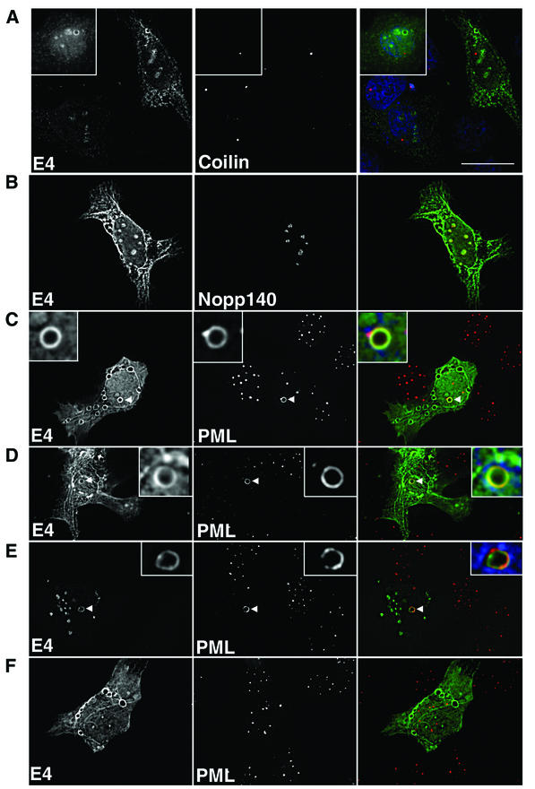
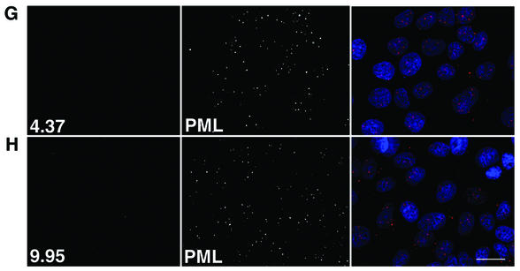
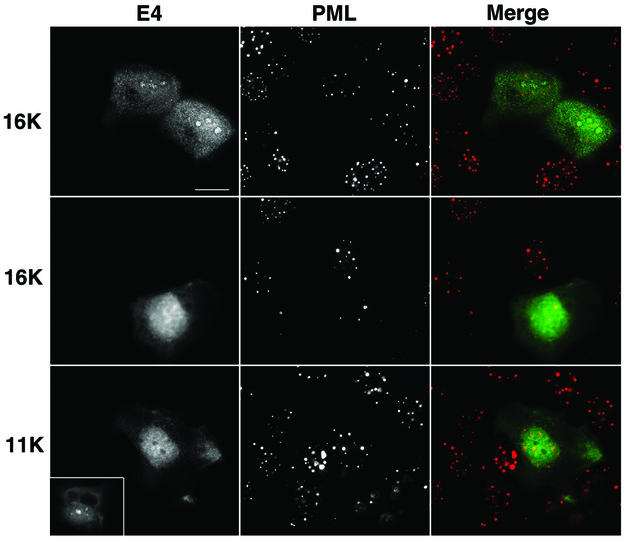
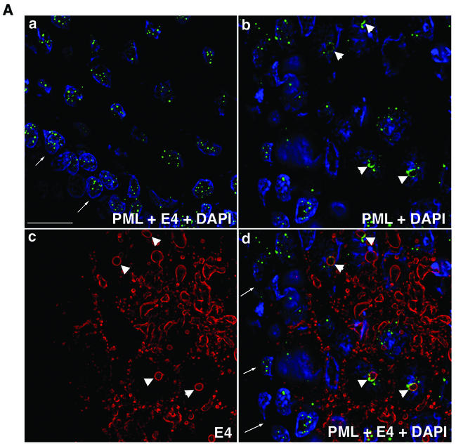
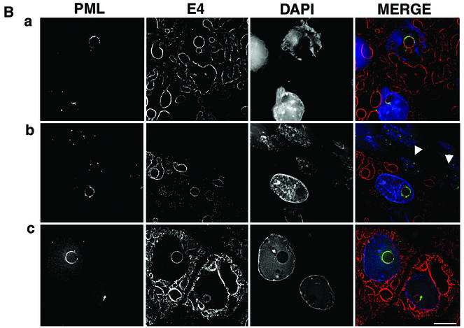
Human papillomavirus type 1 (HPV1) E4 protein is associated with cytoplasmic and nuclear inclusions in productively infected keratinocytes. Here we have used transient expression of HPV1 E4 (also known as E1E4) protein in keratinocytes to reproduce formation of E4 inclusions. Immunofluorescence analysis showed that progressive formation of inclusions correlated with diminished colocalization between E4 and keratin intermediate filaments (IFs). Our results support a model in which the HPV1 E4-keratin IF association is transient, occurring only at an early stage of inclusion formation. We also demonstrate that E4 induces relocation of the promyelocytic leukemia protein (PML) from multiple intranuclear speckles (ND10 bodies) to the periphery of nuclear E4 inclusions and that this activity is specific to full-length E4 protein. Analysis of HPV1-induced warts demonstrated that nuclear PML-E4 inclusions were present in productively infected keratinocytes, indicating that reorganization of PML occurs during the virus's replication cycle. It has been suggested that ND10 bodies are the sites for papillomavirus genome replication and virion assembly. Our finding that E4 induces reorganization of ND10 bodies in vitro and in vivo is further strong evidence that these domains play an important role in the papillomavirus life cycle. This study indicates that HPV1 is analogous to other DNA viruses that disrupt or reorganize ND10 domains, possibly to increase efficiency of virus infection. We hypothesize that HPV1 E4-induced reorganization of PML is necessary for efficient replication of the virus during the virus-producing phase.


Figures

FIG. 1.
FIG. 1.
HPV1 E4 accumulates into in vivo-like inclusion bodies in human keratinocytes. (A) Localization of HPV1 E4 in pcDNA-1E4-transfected SVJD keratinocytes. At 48 h after transfection cells were fixed in 4% paraformaldehyde for 5 min prior to acetone permeabilization (10 min) and stained for E4 (MAb 4.37). Immune complexes were visualized with an anti-mouse Alexa Fluor 488 antibody. HPV1 E4 was associated with multiple inclusion bodies that were both phase dense (c, phase-contrast micrograph of cell shown in panel b; inclusions are indicated with arrowheads) and electron dense (d, electron micrograph showing one of the E4 inclusions formed in an Ad-1E4-infected SVJD cell; bar, 100 nm). Inclusions formed in the cytoplasm and nucleus. A deconvolved z section (0.3 μm) of a cell stained for E4 (e) and counterstained with nuclear stain 4′,6′-diamidino-2-phenylindole (Sigma Chemicals) (f) shows a large intranuclear E4 inclusion (arrowhead). In some cells, E4 was also localized to a cytoplasmic filamentous network (a) and small intranuclear foci that resemble nucleoli (g and h). Bar, 20 μm (6.5 μm in panels e and f). (B) Ad-1E4-infected SVJD keratinocytes at a multiplicity of infection of 25 to 50 were fixed at indicated times p.i. in 4% paraformaldehyde for 1 min prior to permeabilization with acetone. Cells were dual stained for E4 (MAb 4.37) and K18 (MAb CK5) and visualized with anti-mouse immunoglobulin G subclass-specific antibodies conjugated to Alexa 488 and Alexa 594 (Molecular Probes Inc.). Deconvolved z-section images are shown in gray and merged (E4 staining is green, K18 staining is red, and colocalization between red and green stains is shown as yellow). Insets in the top panels show enlargement of the marked area and highlight small E4 inclusions interconnected by E4-K18 filaments. In the middle and bottom panels, arrowheads indicate examples of inclusions that show colocalization between E4 and keratin at the periphery of the E4 inclusion. In the bottom panels arrows show E4 inclusions that are surrounded by keratin staining, but the two antigens do not appear to be colocalized. (C) Ad-1E4-infected SiHa and HaCaT keratinocytes (multiplicity of infection of 50) were fixed 48 and 120 h p.i., respectively. An arrow identifies one of the large inclusions formed in the HaCaT cell. HaCaT cells were a gift from N. Fusenig, University of Heidelberg.
FIG. 1.
FIG. 1.
HPV1 E4 accumulates into in vivo-like inclusion bodies in human keratinocytes. (A) Localization of HPV1 E4 in pcDNA-1E4-transfected SVJD keratinocytes. At 48 h after transfection cells were fixed in 4% paraformaldehyde for 5 min prior to acetone permeabilization (10 min) and stained for E4 (MAb 4.37). Immune complexes were visualized with an anti-mouse Alexa Fluor 488 antibody. HPV1 E4 was associated with multiple inclusion bodies that were both phase dense (c, phase-contrast micrograph of cell shown in panel b; inclusions are indicated with arrowheads) and electron dense (d, electron micrograph showing one of the E4 inclusions formed in an Ad-1E4-infected SVJD cell; bar, 100 nm). Inclusions formed in the cytoplasm and nucleus. A deconvolved z section (0.3 μm) of a cell stained for E4 (e) and counterstained with nuclear stain 4′,6′-diamidino-2-phenylindole (Sigma Chemicals) (f) shows a large intranuclear E4 inclusion (arrowhead). In some cells, E4 was also localized to a cytoplasmic filamentous network (a) and small intranuclear foci that resemble nucleoli (g and h). Bar, 20 μm (6.5 μm in panels e and f). (B) Ad-1E4-infected SVJD keratinocytes at a multiplicity of infection of 25 to 50 were fixed at indicated times p.i. in 4% paraformaldehyde for 1 min prior to permeabilization with acetone. Cells were dual stained for E4 (MAb 4.37) and K18 (MAb CK5) and visualized with anti-mouse immunoglobulin G subclass-specific antibodies conjugated to Alexa 488 and Alexa 594 (Molecular Probes Inc.). Deconvolved z-section images are shown in gray and merged (E4 staining is green, K18 staining is red, and colocalization between red and green stains is shown as yellow). Insets in the top panels show enlargement of the marked area and highlight small E4 inclusions interconnected by E4-K18 filaments. In the middle and bottom panels, arrowheads indicate examples of inclusions that show colocalization between E4 and keratin at the periphery of the E4 inclusion. In the bottom panels arrows show E4 inclusions that are surrounded by keratin staining, but the two antigens do not appear to be colocalized. (C) Ad-1E4-infected SiHa and HaCaT keratinocytes (multiplicity of infection of 50) were fixed 48 and 120 h p.i., respectively. An arrow identifies one of the large inclusions formed in the HaCaT cell. HaCaT cells were a gift from N. Fusenig, University of Heidelberg.
FIG. 2.
FIG. 2.
Subcellular distribution of HPV1 E4 in human fibroblasts. (A and B) Ad-1E4-infected human primary fibroblasts (multiplicity of infection of 50) were fixed 48 h p.i. Cells stained with an anti-HPV1 E4 MAb, 4.37, showed E4 localized to small cytoplasmic and intranuclear inclusions (indicated by arrowheads) that appeared identical to those formed in keratinocytes (inset, phase-contrast image of part of cell shown in panel A, which shows inclusions to be phase dense). E4 staining also formed a cytoplasmic network (B) that coaligned with actin stress fibers (C) . (D to G) Dual staining of cells infected with Ad-1E4Δ2-15 (D and E) or Ad-1E4Δ10-14 (F and G) showed that the mutant proteins associated with the actin cytoskeleton (D and F, anti-HPV1 MAb 4.37; E and G, phalloidin). Bar, 20 μm in panels A to E and 13 μm in panels F and G.
FIG. 3.
FIG. 3.
HPV2 E4 colocalizes to keratin IFs and induces collapse of the keratin matrix into a fibrous bundle. SVJD cells were transfected with pcDNA-2E4, fixed 48 h p.i., and dual stained with anti-HPV2 E4 MAb IF10 and K18 MAb. Note that the keratin cytoskeleton in neighboring E4-negative cells remains intact; one is shown by the arrow. Bar, 20 μm.
FIG. 4.
FIG. 4.
Subnuclear topology of HPV1 E4 in SVJD keratinocytes. pcDNA-1E4-transfected SVJD cells were fixed at 48 h in 4% paraformaldehyde for 5 min (A to D and F) or 1 min (E) and permeabilized in acetone. Cells were dual stained for E4 (MAb 4.37) and cellular factors of coilin bodies (A, coilin), nucleoli (B, Nopp140), and ND10 bodies (C to F; PML, rabbit anti-PML antibody). Deconvolved z sections are shown as individual images (gray) and merged (right panels). In merged images, E4 is green; coilin, Nopp140, and PML are red; and where shown 4′,6′-diamidino-2-phenylindole staining of nuclei is blue. Yellow in merged images indicates colocalization between green and red colors. (C to E) Note that in cells containing E4 inclusion bodies PML was reorganized to the periphery of the intranuclear E4 inclusion. The insets in panels C to E are enlargements of the inclusion bodies indicated by arrowheads. PML was partly colocalized with E4. (E) PML-E4 inclusions were also observed in cells that had been fixed differently and stained with a different anti-PML antibody (MAb PG-M3). (F) Note that reorganization of PML did not occur in cells that contain only cytoplasmic E4 inclusions. (G and H) Mock-transfected cells dual stained with 4.37 (G) or 9.95 (H) and PML. Bar, 20 μm (insets, 5.5 μm).
FIG. 4.
FIG. 4.
Subnuclear topology of HPV1 E4 in SVJD keratinocytes. pcDNA-1E4-transfected SVJD cells were fixed at 48 h in 4% paraformaldehyde for 5 min (A to D and F) or 1 min (E) and permeabilized in acetone. Cells were dual stained for E4 (MAb 4.37) and cellular factors of coilin bodies (A, coilin), nucleoli (B, Nopp140), and ND10 bodies (C to F; PML, rabbit anti-PML antibody). Deconvolved z sections are shown as individual images (gray) and merged (right panels). In merged images, E4 is green; coilin, Nopp140, and PML are red; and where shown 4′,6′-diamidino-2-phenylindole staining of nuclei is blue. Yellow in merged images indicates colocalization between green and red colors. (C to E) Note that in cells containing E4 inclusion bodies PML was reorganized to the periphery of the intranuclear E4 inclusion. The insets in panels C to E are enlargements of the inclusion bodies indicated by arrowheads. PML was partly colocalized with E4. (E) PML-E4 inclusions were also observed in cells that had been fixed differently and stained with a different anti-PML antibody (MAb PG-M3). (F) Note that reorganization of PML did not occur in cells that contain only cytoplasmic E4 inclusions. (G and H) Mock-transfected cells dual stained with 4.37 (G) or 9.95 (H) and PML. Bar, 20 μm (insets, 5.5 μm).
FIG. 5.
FIG. 5.
HPV1 E4 proteins that lack N-terminal sequences do not induce reorganization of PML in keratinocytes. SVJD keratinocytes were transfected for 48 h with pcDNA-1E4Δ2-15 (top and middle panels) and pcDNA-1E4Δ2-58 (bottom panels). These plasmids express proteins that closely resemble the truncated E4 proteins, 16K and 11K, expressed in HPV1-induced warts (45). Cells were dual stained for E4 (MAb 4.37, top and middle panels; p1p7 antibody, bottom panels) and PML (rabbit anti-PML antibody). Note that the Δ2-58 protein lacks epitopes of anti-HPV1 E4 MAbs and therefore was recognized with a rat polyclonal antibody, p1p7 (18). In merged images, E4 is green and PML is red. Truncated proteins do not assemble into inclusions in SVJD cells, and PML is localized to multiple speckles, a similar distribution as that of PML in adjacent nontransfected cells. However, cells expressing high levels of 16K protein often contain a low number of PML-stained speckles (a typical example is shown in the middle panels). Note that 16K, but not 11K, E4 is localized to nucleoli (compare top with bottom panels). The inset in the bottom panel shows a 16K-expressing cell stained with p1p7 to show that this antibody is able to recognize nucleolar E4. Bar, 20 μm (inset, 25 μm).
FIG. 6.
FIG. 6.
PML is redistributed to intranuclear E4 inclusions in HPV1-induced warts. (A) Tissue sections of wart were dual stained for E4 (MAb 9.95; E4 staining is now shown in red) and PML (rabbit antibody, green), and nuclei were counterstained with 4′,6′-diamidino-2-phenylindole (DAPI) (blue). (a) Merged deconvolved z sections of area of wart negative for E4 staining. Note that PML was localized to multiple intranuclear speckles in basal cells (arrows indicate basal layer) and suprabasal cells. (b to d) PML distribution in area of wart expressing E4. Deconvolved z-section images are shown as PML and DAPI merged (b); E4 alone (c); and E4, PML, and DAPI merged (d). In suprabasal cells, PML was reorganized from multiple speckles to ring structures (b) that are localized to intranuclear E4 inclusions (d; arrows indicate basal cell layer). Arrowheads indicate PML-E4 inclusions. Bar, 20 μm. (B) Reorganization of PML to E4 inclusions was identical to that observed for E4-expressing cultured keratinocytes. Deconvolved z sections are shown as individual images (gray) and merged (E4, red; PML, green; DAPI, blue). PML either nearly completely surrounds the E4 inclusion (a and c) or accumulates predominantly to one side (b). PML shows partial colocalization with E4 (depicted as yellow color in merged images). Note that in panel b the E4-positive cell immediately above the basal layer (indicated by arrowheads) contains a PML-E4 inclusion and that PML distribution is normal in basal cells. Note that the wart section in panel a is stained with MAb 9.95 and that wart sections in panels b and c are stained with anti-E4 MAb 1D11. MAb 1D11 recognizes only full-length E4 (E1^E4) protein. Bar, 10 μm.
FIG. 6.
FIG. 6.
PML is redistributed to intranuclear E4 inclusions in HPV1-induced warts. (A) Tissue sections of wart were dual stained for E4 (MAb 9.95; E4 staining is now shown in red) and PML (rabbit antibody, green), and nuclei were counterstained with 4′,6′-diamidino-2-phenylindole (DAPI) (blue). (a) Merged deconvolved z sections of area of wart negative for E4 staining. Note that PML was localized to multiple intranuclear speckles in basal cells (arrows indicate basal layer) and suprabasal cells. (b to d) PML distribution in area of wart expressing E4. Deconvolved z-section images are shown as PML and DAPI merged (b); E4 alone (c); and E4, PML, and DAPI merged (d). In suprabasal cells, PML was reorganized from multiple speckles to ring structures (b) that are localized to intranuclear E4 inclusions (d; arrows indicate basal cell layer). Arrowheads indicate PML-E4 inclusions. Bar, 20 μm. (B) Reorganization of PML to E4 inclusions was identical to that observed for E4-expressing cultured keratinocytes. Deconvolved z sections are shown as individual images (gray) and merged (E4, red; PML, green; DAPI, blue). PML either nearly completely surrounds the E4 inclusion (a and c) or accumulates predominantly to one side (b). PML shows partial colocalization with E4 (depicted as yellow color in merged images). Note that in panel b the E4-positive cell immediately above the basal layer (indicated by arrowheads) contains a PML-E4 inclusion and that PML distribution is normal in basal cells. Note that the wart section in panel a is stained with MAb 9.95 and that wart sections in panels b and c are stained with anti-E4 MAb 1D11. MAb 1D11 recognizes only full-length E4 (E1^E4) protein. Bar, 10 μm.

Similar articles

Cited by

References

    1. Adamson, A. L., and S. Kenney. 2001. Epstein-Barr virus immediate-early protein BZLF1 is SUMO-1 modified and disrupts promyelocytic leukemia bodies. J. Virol. 75:2388-2399. - PMC - PubMed
    1. Ahn, J. H., and G. S. Hayward. 1997. The major immediate-early proteins IE1 and IE2 of human cytomegalovirus colocalize with and disrupt PML-associated nuclear bodies at early times in infected permissive cells. J. Virol. 71:4599-4613. - PMC - PubMed
    1. Almeida, F., R. Saffrich, W. Ansorge, and M. Carmo-Fonseca. 1998. Microinjection of anti-coilin antibodies affects the structure of coiled bodies. J. Cell Biol. 142:899-912. - PMC - PubMed
    1. Ashmole, I., P. H. Gallimore, and S. Roberts. 1998. Identification of conserved hydrophobic C-terminal residues of the human papillomavirus type 1 E1^E4 protein necessary for E4 oligomerisation in vivo. Virology 240:221-231. - PubMed
    1. Bell, P., P. M. Lieberman, and G. G. Maul. 2000. Lytic but not latent replication of Epstein-Barr virus is associated with PML and induces sequential release of nuclear domain 10 proteins. J. Virol. 74:11800-11810. - PMC - PubMed

Publication types