

doi: 10.1128/jvi.77.1.726-731.2003.
Human and simian immunodeficiency virus capsid proteins are major viral determinants of early, postentry replication blocks in simian cells
Affiliations
- PMID: 12477877
- PMCID: PMC140632
- DOI: 10.1128/jvi.77.1.726-731.2003
Item in Clipboard
Human and simian immunodeficiency virus capsid proteins are major viral determinants of early, postentry replication blocks in simian cells
J Virol.
2003 Jan.
Abstract
The cells of most Old World monkey species exhibit early, postentry restrictions on infection by human immunodeficiency virus type 1 (HIV-1) but not by simian immunodeficiency virus of macaques (SIV(mac)). Conversely, SIV(mac), but not HIV-1, infection is blocked in most New World monkey cells. By using chimeric HIV-1/SIV(mac) viruses capable of a single round of infection, we demonstrated that a major viral determinant of this restriction is the capsid (CA) protein. The efficiency of early events following HIV-1 and SIV(mac) entry is apparently determined by the interaction of the incoming viral CA and species-specific host factors.
Figures

HIV-1/SIVmac recombinants. The name of the construct reflects the HIV-1 or SIV proviral backbone, and the names in parentheses designate the source of the CA or CA-p2 sequences (H = HIV-1; S = SIVmac). The amino-terminal portions of the Gag polyproteins of the recombinant viruses used in this study are shown beneath the wild-type HIV-1 (white) and SIVmac239 (black) sequences. The locations of the matrix (MA), CA, p2, and NC proteins are shown. The numbers represent the boundaries of the HIV-1 CA sequence that was replaced by the SIVmac sequence. The locations of the N- and C-terminal domains of the CA proteins are shown. The asterisk represents the approximate location of a threonine-to-cysteine change at residue 58 of the HIV-1 CA protein.
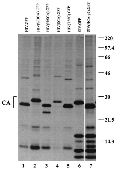
Virion proteins produced by HIV-1/SIVmac chimeras. 293T cells were transfected with plasmids HIV.GFP (lane 1), HIV(S/HCA).GFP (lane 2), HIV(H/SCA).GFP (lane 3), HIV(SCA).GFP (lane 4), HIV(T58C).GFP (lane 5), SIV.GFP (lane 6), and SIV(HCA-p2).GFP (lane 7). Following transfection, the cells were labeled with [35S]methionine. Virions released into the cell supernatants were pelleted through 20% sucrose and resuspended in radioimmunoprecipitation assay buffer before analysis by sodium dodecyl sulfate-12.5% polyacrylamide gel electrophoresis and autoradiography. The location of the CA proteins is shown on the left. The values on the right are molecular sizes in kilodaltons.
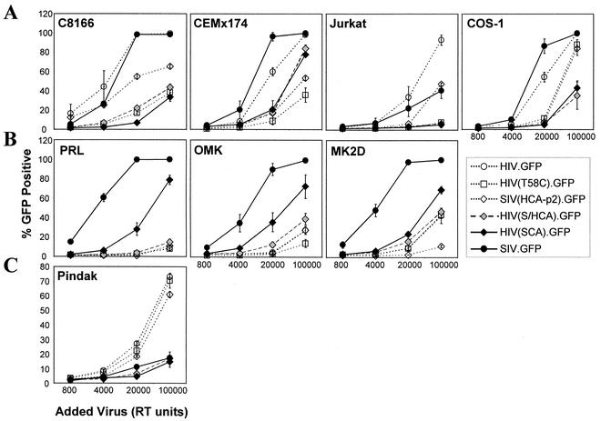
Infection of cells by HIV-1/SIVmac recombinants. Supernatants containing the indicated single-round HIV-1 and SIVmac vectors, which encode GFP, were assessed for reverse transcriptase (RT) activity. The indicated number of RT units of virus was added to cells, which were incubated for 3 days prior to fluorescence-activated cell sorting analysis for GFP expression. The cells in the top row (A) are permissive for both HIV-1 and SIVmac, the cells in the second row (B) are permissive for SIVmac but restricted for HIV-1, and the cells in panel C are permissive for HIV-1 but restricted for SIVmac. The symbols and lines used for the viruses are as follows: viruses with HIV-1 CA sequences, open symbols and dotted lines; viruses with SIVmac CA sequences, black symbols and solid lines; virus with CA sequences derived from SIVmac and HIV-1, gray symbol and dashed line.
References
-
- Alkhatib, G., C. Combadiere, C. C. Broder, Y. Feng, P. E. Kennedy, P. M. Murphy, and E. A. Berger. 1996. CC CKR5: a RANTES, MIP-1α, MIP-1β receptor as a fusion cofactor for macrophage-tropic HIV-1. Science 272:1955-1958. - PubMed
-
- Balzarini, J., E. de Clercq, and K. Uberla. 1997. SIV/HIV-1 hybrid virus expressing the reverse transcriptase gene of HIV-1 remains sensitive to HIV-1-specific reverse transcriptase inhibitors after passage in rhesus macaques. J. AIDS Hum. Retrovirol. 15:1-4. - PubMed
-
- Barre-Sinoussi, F., J. C. Chermann, F. Rey, M. T. Nugeyre, S. Chamaret, J. Gruest, C. Dauguet, C. Axler-Blin, F. Vezinet-Brun, C. Rouzioux, W. Rozenbaum, and L. Montagnier. 1983. Isolation of a T-lymphotropic retrovirus from a patient at risk for acquired immune deficiency syndrome (AIDS). Science 220:868-871. - PubMed
Publication types
MeSH terms
Grants and funding
LinkOut - more resources
Full Text Sources
Other Literature Sources
