

Time between onset of apoptosis and release of nucleosomes from apoptotic cells: putative implications for systemic lupus erythematosus
- PMID: 12480662
- PMCID: PMC1754285
- DOI: 10.1136/ard.62.1.10
Time between onset of apoptosis and release of nucleosomes from apoptotic cells: putative implications for systemic lupus erythematosus
Abstract
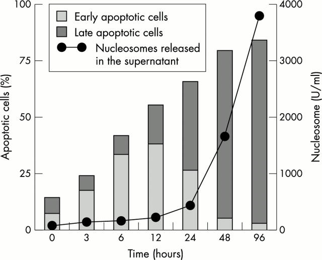
Objective: To investigate the kinetics of nucleosome leakage from apoptotic cells in an in vitro system and extrapolate the results to autoimmune disease, in particular systemic lupus erythematosus.
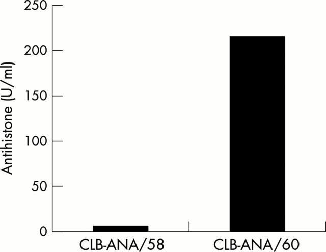
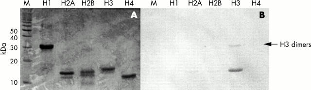
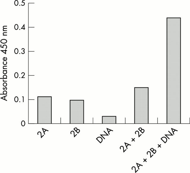
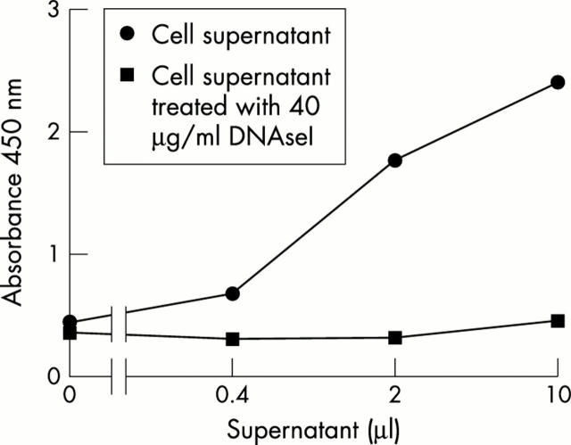
Methods: A sensitive nucleosome enzyme linked immunosorbent assay (ELISA) was developed, using a monoclonal antibody (mAb) against histone 3 and an mAb against nucleosomes. Nucleosome release during apoptotic cell death was studied in Jurkat cells. AnnexinV binding (early apoptosis) and propidium iodide positivity (late apoptosis) of the cells were compared with nucleosome release at different times after apoptosis induction.
Results: Nucleosomes appeared in culture supernatant of Jurkat cells 24 to 48 hours after apoptosis induction, when the cells had been late apoptotic for more than 12 hours.
Conclusion: Nucleosomes are released from late apoptotic Jurkat cells, with a 12 hour delay from the appearance of AnnexinV binding cells. This result suggests that in vivo scavenger mechanisms have 12 hours to remove apoptotic material from the circulation.
Figures
Comment in
-
Defective waste disposal: does it induce autoantibodies in SLE?Ann Rheum Dis. 2003 Jan;62(1):1-3. doi: 10.1136/ard.62.1.1-a. Ann Rheum Dis. 2003. PMID: 12480659 Free PMC article. No abstract available.
References
Publication types
MeSH terms
Substances
LinkOut - more resources
Full Text Sources
Other Literature Sources
Medical
