. 2002 Dec 24;99(26):16742-7.
doi: 10.1073/pnas.262663499. Epub 2002 Dec 12.

A structural model for Alzheimer's beta -amyloid fibrils based on experimental constraints from solid state NMR

Affiliations

A structural model for Alzheimer's beta -amyloid fibrils based on experimental constraints from solid state NMR

Aneta T Petkova et al. Proc Natl Acad Sci U S A. .

Abstract

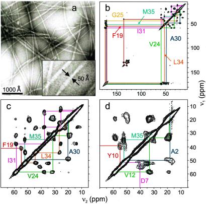
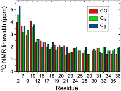
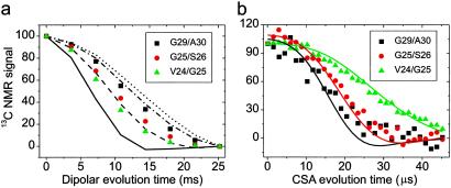
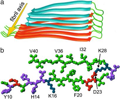
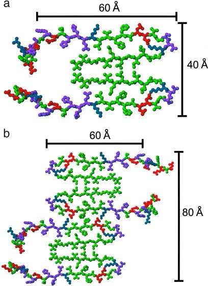
We present a structural model for amyloid fibrils formed by the 40-residue beta-amyloid peptide associated with Alzheimer's disease (Abeta(1-40)), based on a set of experimental constraints from solid state NMR spectroscopy. The model additionally incorporates the cross-beta structural motif established by x-ray fiber diffraction and satisfies constraints on Abeta(1-40) fibril dimensions and mass-per-length determined from electron microscopy. Approximately the first 10 residues of Abeta(1-40) are structurally disordered in the fibrils. Residues 12-24 and 30-40 adopt beta-strand conformations and form parallel beta-sheets through intermolecular hydrogen bonding. Residues 25-29 contain a bend of the peptide backbone that brings the two beta-sheets in contact through sidechain-sidechain interactions. A single cross-beta unit is then a double-layered beta-sheet structure with a hydrophobic core and one hydrophobic face. The only charged sidechains in the core are those of D23 and K28, which form salt bridges. Fibrils with minimum mass-per-length and diameter consist of two cross-beta units with their hydrophobic faces juxtaposed.


Figures

Fig 1.
Fig 1.
(a) Transmission electron microscope images of negatively stained amyloid fibrils after 14-day incubation of a 0.5 mM Aβ1–40 solution. A 3× expansion (Inset) shows fibrils with the smallest diameters observed. (b) 2D 13C-13C chemical shift correlation spectrum of Aβ1–40 fibril sample SU7, showing resonance assignment paths for the seven uniformly 15N- and 13C-labeled residues in this sample. (c) Expansion of the aliphatic region of the 2D spectrum of SU7. (d) Aliphatic region of the 2D 13C-13C chemical shift correlation spectrum of Aβ1–40 fibril sample SU6.
Fig 2.
Fig 2.
13C NMR linewidths for CO, Cα, and Cβ sites in Aβ1–40 fibrils, determined from 2D solid state NMR spectra as in Fig. 1. Linewidths of 2.5 ppm or less indicate well-ordered conformations. Larger linewidths in the N-terminal segment indicate structural disorder.
Fig 3.
Fig 3.
Solid state NMR data on DLn Aβ1–40 fibril samples with 13C labels at the indicated backbone carbonyl sites. These data constrain the φ and ψ angles of the second labeled residue. (a) fpRFDR-CT data and simulations for φ = 40° (solid line), 80° (dashed line), 120° (dot-dashed line), and 160° (dotted line). Simulations are scaled and baseline-corrected to match the first and last experimental data points. (b) DQCSA data and simulations for φ, ψ = −70°, −40° (green); 70°, −65° (red); and −165°, 135° (black).
Fig 4.
Fig 4.
Structural model for Aβ1–40 fibrils, consistent with solid state NMR constraints on the molecular conformation and intermolecular distances and incorporating the cross-β motif common to all amyloid fibrils. Residues 1–8 are considered fully disordered and are omitted. (a) Schematic representation of a single molecular layer, or cross-β unit. The yellow arrow indicates the direction of the long axis of the fibril, which coincides with the direction of intermolecular backbone hydrogen bonds. The cross-β unit is a double-layered structure, with in-register parallel β-sheets formed by residues 12–24 (orange ribbons) and 30–40 (blue ribbons). (b) Central Aβ1–40 molecule from the energy-minimized, five-chain system, viewed down the long axis of the fibril. Residues are color-coded according to their sidechains as hydrophobic (green), polar (magenta), positive (blue), or negative (red).
Fig 5.
Fig 5.
(a) Cross section of an Aβ1–40 fibril with the minimal MPL indicated by scanning transmission electron microscopy (13, 29), formed by juxtaposing the hydrophobic faces of two cross-β units from Fig. 4. Residues 1–8 are included with randomly assigned conformations. (b) Possible mode of lateral association to generate fibrils with greater MPL and greater cross-sectional dimensions.

Comment in

Similar articles

Cited by

References

    1. Sipe J. D. (1992) Annu. Rev. Biochem. 61, 947-975. - PubMed
    1. Sunde M., Serpell, L. C., Bartlam, M., Fraser, P. E., Pepys, M. B. & Blake, C. C. F. (1997) J. Mol. Biol. 273, 729-739. - PubMed
    1. Inouye H., Fraser, P. E. & Kirschner, D. A. (1993) Biophys. J. 64, 502-519. - PMC - PubMed
    1. Glenner G. G. & Wong, C. W. (1984) Biochem. Biophys. Res. Commun. 120, 885-890. - PubMed
    1. Iwatsubo T., Odaka, A., Suzuki, N., Mizusawa, H., Nukina, N. & Ihara, Y. (1994) Neuron 13, 45-53. - PubMed

LinkOut - more resources