. 2002 Dec 24;99(26):16770-5.
doi: 10.1073/pnas.222679399. Epub 2002 Dec 12.

Low intracellular zinc induces oxidative DNA damage, disrupts p53, NFkappa B, and AP1 DNA binding, and affects DNA repair in a rat glioma cell line

Affiliations

Low intracellular zinc induces oxidative DNA damage, disrupts p53, NFkappa B, and AP1 DNA binding, and affects DNA repair in a rat glioma cell line

Emily Ho et al. Proc Natl Acad Sci U S A. .

Abstract

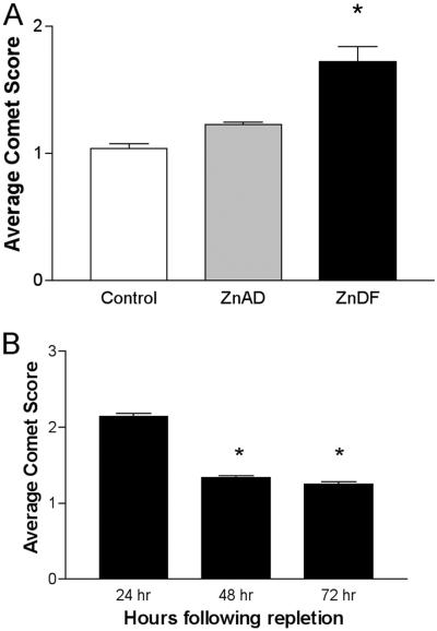
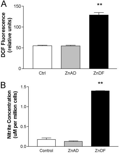
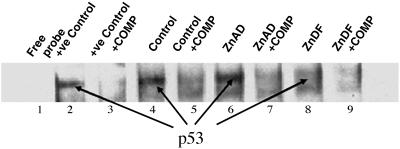
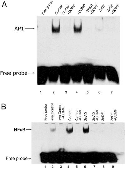
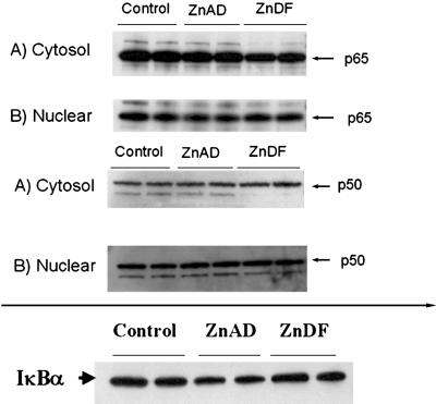
Approximately 10% of the U.S. population ingests <50% of the current recommended daily allowance for zinc. We investigate the effect of zinc deficiency on DNA damage, expression of DNA-repair enzymes, and downstream signaling events in a cell-culture model. Low zinc inhibited cell growth of rat glioma C6 cells and increased oxidative stress. Low intracellular zinc increased DNA single-strand breaks (comet assay). Zinc-deficient C6 cells also exhibited an increase in the expression of the zinc-containing DNA-repair proteins p53 and apurinic endonuclease (APE). Repletion with zinc restored cell growth and reversed DNA damage. APE is a multifunctional protein that not only repairs DNA but also controls DNA-binding activity of many transcription factors that may be involved in cancer progression. The ability of the transcription factors p53, nuclear factor kappaB, and activator protein 1 (AP1) to bind to consensus DNA sequences was decreased markedly with zinc deficiency, as assayed by electrophoretic mobility-shift assays. Thus, low intracellular zinc status causes oxidative DNA damage and induces DNA-repair protein expression, but binding of p53 and important downstream signals leading to proper DNA repair are lost without zinc.


Figures

Fig 1.
Fig 1.
Cellular copper, iron, and zinc concentrations in ZnDF C6 cells. Mineral levels were determined by inductively coupled plasma/absorption emission spectrometry as described in Materials and Methods. For each group, cells were fed control-DMEM (Control), ZnAD-DMEM (ZnAD), or ZnDF-DMEM (ZnDF) for a 5-day period. All samples are mean ± SE (n = 6). Significant differences between means were determined by one-way ANOVA. **, P < 0.01.
Fig 2.
Fig 2.
Zinc deficiency induces single-strand breaks in C6 cells. (A) C6 cells were grown in control, ZnAD, or ZnDF medium for 5 days. Each sample is representative of an average mean comet score ± SE of four individual slides per treatment. (B) ZnDF cells were repleted with ZnAD medium. On each slide, 50 comets were scored blindly for tail moment. *, P < 0.05.
Fig 3.
Fig 3.
Zinc deficiency increases oxidant production and NO formation in ZnDF C6 cells. (A) DCFH probe was used to monitor oxidant production in C6 cells. DCFH was added to C6 cells grown in control, ZnAD, or ZnDF medium for 5 days. (B) Nitrite production was used as an indirect measure of NO production. Nitrite levels were assessed in media after 5 days in culture in control, ZnAD, or ZnDF medium. **, P < 0.01.
Fig 4.
Fig 4.
Zinc deficiency increases expression of p53 and APE. p53 and APE expression were determined by Western blot analysis in cells grown in control, ZnAD, or ZnDF medium for 5 days. SDS/PAGE was performed as outlined in Materials and Methods. Blots were stripped and blotted for actin to confirm each protein loading (data not shown). Each bar is representative of mean ± SE (n = 3). *, P < 0.05.
Fig 5.
Fig 5.
p53 is compromised in ZnDF C6 cells. EMSAs were performed with p53, AP1, or NFκB oligonucleotide probes to examine binding in nuclear extracts from cells grown in control, ZnAD, and ZnDF medium for 5 days. Lane 1 contains a free oligonucleotide probe, and lane 2 contains a positive control using HeLa cells (Promega) that contains active p53 binding. Lanes 3, 5, 7, and 9 represent nuclear extracts incubated with specific competitor (COMP, unlabeled p53 oligonucleotide) to confirm specificity of p53 binding. +ve, positive.
Fig 6.
Fig 6.
AP1 and NFκB binding is compromised in ZnDF C6 cells. (A) AP1 binding: lane 1 contains a free oligonucleotide probe. (B) NFκB binding: lane 1 contains a free oligonucleotide probe, and lane 2 contains a positive control using HeLa cells (Promega) that contains active NFκB. Lanes 3, 5, 7, and 9 represent nuclear extracts incubated with unlabeled oligonucleotide (COMP) to confirm specificity of binding. +ve, positive.
Fig 7.
Fig 7.
Zinc deficiency has no effect on nuclear translocation of NFκB subunits p50 or p65 or IκBα degradation. Western blot analysis was used to determine protein levels of p50, p65, and IκBα in nuclear and cytoplasmic fractions as described in Materials and Methods. SDS/PAGE was performed in nuclear and cytoplasmic fractions from cells grown in control, ZnAD, or ZnDF medium. Blots were stripped and blotted for actin to confirm each protein loading (data not shown). Results are representative of two individual experiments.

References

    1. Ames B. N. & Wakimoto, P. (2002) Nat. Rev. Cancer 2, 694-704. - PubMed
    1. Ames B. N. (2001) Mutat. Res. 475, 7-20. - PubMed
    1. Ames B. N. & Gold, L. S. (2000) Mutat. Res. 447, 3-13. - PubMed
    1. Blount B. C., Mack, M. M., Wehr, C. M., MacGregor, J. T., Hiatt, R. A., Wang, G., Wickramasinghe, S. N., Everson, R. B. & Ames, B. N. (1997) Proc. Natl. Acad. Sci. USA 94, 3290-3295. - PMC - PubMed
    1. Elliott R. L., Elliott, M. C., Wang, F. & Head, J. F. (1993) Ann. N.Y. Acad. Sci. 698, 159-166. - PubMed

Publication types