Comparative Study
. 2002 Dec;130(4):1871-82.
doi: 10.1104/pp.008573.

Comparative molecular and functional analyses of the tobacco cyclin-dependent kinase inhibitor NtKIS1a and its spliced variant NtKIS1b

Affiliations
Comparative Study

Comparative molecular and functional analyses of the tobacco cyclin-dependent kinase inhibitor NtKIS1a and its spliced variant NtKIS1b

Sophie Jasinski et al. Plant Physiol. 2002 Dec.

Abstract

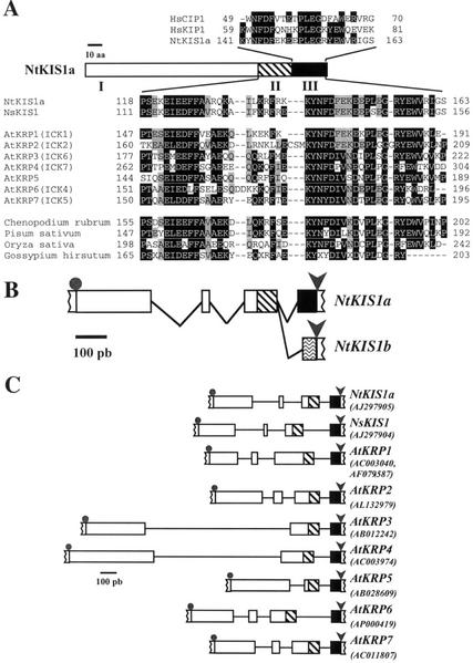
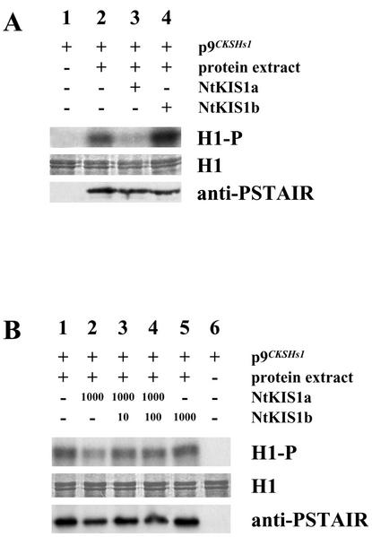
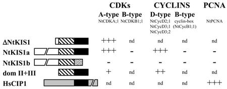
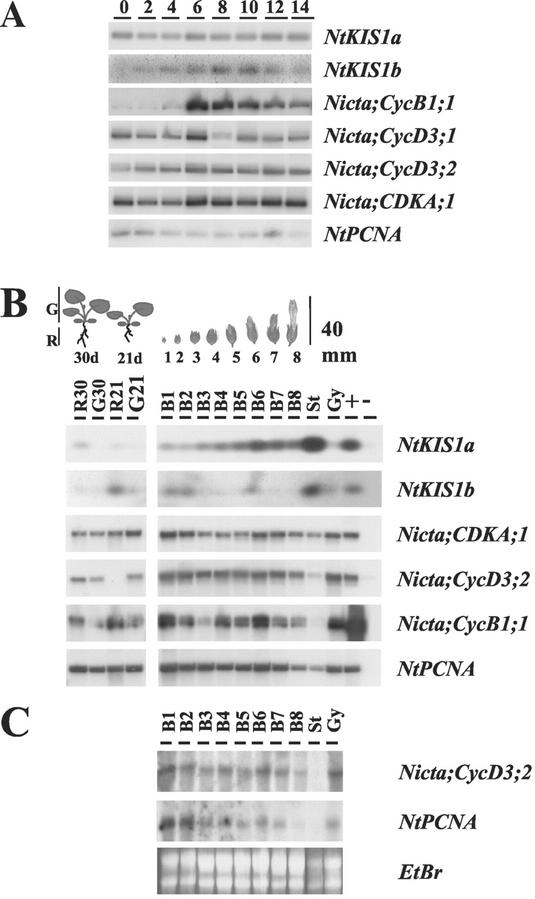
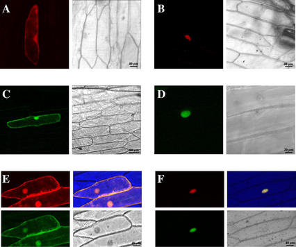
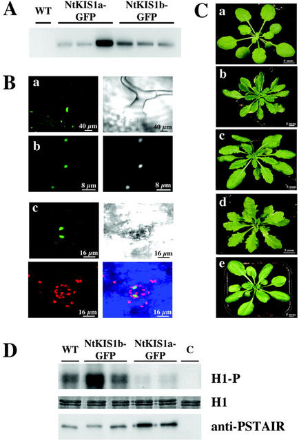
In all eukaryotes, cell cycle progression is controlled by cyclin-dependent kinases (CDKs) whose activity is regulated at several levels including inhibition by CDK inhibitors. Here, we report a comparative molecular and functional analysis of the tobacco (Nicotiana tomentosiformis) CDK inhibitor, NtKIS1a, and its spliced variant, NtKIS1b. The C-terminal end of NtKIS1a shares strong sequence similarity with mammalian CIP/KIP inhibitors, which is not the case for NtKIS1b. Consistent with this, NtKIS1a but not NtKIS1b inhibits in vitro the kinase activity of CDK/cyclin complexes, and tobacco (Nicotiana tabacum) D-type cyclins and an A-type CDK are NtKIS1a, but not NtKIS1b, interacting partners. Although both NtKIS1a and NtKIS1b transcripts are mainly found in flowers and more precisely in stamens, NtKIS1b transcript levels are cell cycle regulated, whereas those of NtKIS1a remain constant during the cell cycle. NtKIS1a and NtKIS1b fused to fluorescent proteins are localized in the nucleus when transiently expressed in onion epidermal cells. Furthermore, there is no competition for their nuclear localization when they are simultaneously overexpressed. In vitro competition toward CDK kinase activity suggests that NtKIS1b is a strong competitor of NtKIS1a. Arabidopsis plants overexpressing NtKIS1a-green fluorescent protein (GFP) or NtKIS1b-GFP fusion proteins were obtained. In these plants, the fusion proteins are still localized in the nucleus. Interestingly, NtKIS1a-GFP-overexpressing plants display strong morphological modifications and a reduced CDK kinase activity, whereas NtKIS1b-GFP-overexpressing plants display a wild-type phenotype including a wild-type CDK kinase activity. Our results strongly suggest that the inhibition of the kinase activity is responsible for the phenotypic modifications.


Figures

Figure 1
Figure 1
NtKIS1 sequence analysis. A, The amino acid sequence deduced from NtKIS1a is schematically represented with its three domains: domain I (residues 1–117; white box), domain II (residues 118–140; hatched box), and domain III (residues 141–163; black box; also in B and C). Alignment of the domain III with human CIP/KIP inhibitors is shown above (HsCIP1: L25610; HsKIP1: U10906). Alignment of domains II+III with plant-related proteins is shown below. NsKIS1 corresponds to the polypeptide deduced from the GenScan-predicted open reading frame of N. sylvestris genomic sequence (http://bioweb.pasteur.fr/seqanal/interfaces/genscan.html). AtKRP1 to AtKRP7 correspond to the Arabidopsis polypeptides deduced from cDNA sequences. Correspondences with ICKs are given in brackets. C. rubrum, pea, and rice correspond to polypeptides deduced from cDNA sequences (AJ002173, AB029483, and AC069145). Cotton corresponds to the polypeptide deduced from an incomplete EST (AI728644). Identical or similar amino acids present in more than 50% and present in 30% to 50% of the proteins are highlighted, respectively, in black and gray. B, The exon-intron organization of the N. tomentosiformis NtKIS1 genomic sequence, which results from the comparison with NtKIS1a and NtKIS1b cDNAs, is schematically represented with a potential alternative splicing of the third intron (exons, boxes; introns, lines). Gray dots and arrows indicate, respectively, start and stop codons (also in C). Waved box represents the fourth exon in NtKIS1b different from NtKIS1a domain III defined above. C, The exon-intron organizations deduced from the different genomic sequences are compared. Accession numbers are indicated.
Figure 2
Figure 2
In vitro effect of purified recombinant NtKIS1a and NtKIS1b proteins on the kinase activity of BY-2 CDK/cyclin complexes. A, Histone H1 phosphorylation is shown (H1-P). Protein extract of 24-h refreshed BY-2 cells was added (+, lanes 2–4) or not (−, lane 1) to p9CKSHs1 beads to purify CDK/cyclin complexes. Histone H1 (25 μg) was added in all lanes (H1). Five hundred nanograms of NtKIS1a (lane 3), 500 ng of NtKIS1b (lane 4), or buffer (lanes 1 and 2) was further added. Histone H1K activity was monitored as described in “Materials and Methods.” Proteins bound to p9CKSHs1 beads were recovered and immunoblotted with an antibody raised against the conserved CDK PSTAIR motif (anti-PSTAIR). B, Histone H1 phosphorylation is shown (H1-P). Protein extract of 24-h refreshed BY-2 cells was added (+, lanes 1–5) or not (−, lane 6) to p9CKSHs1 beads to purify CDK/cyclin complexes. Histone H1 (25 μg) was added in all lanes (H1). One thousand nanograms of NtKIS1a (lanes 2–4), 10 ng of NtKIS1b (lane 3), 100 ng of NtKIS1b (lane 4), 1,000 ng of NtKIS1b (lane 5), or buffer (lanes 1 and 6) was further added. Histone H1K activity was monitored as described in “Materials and Methods.” Proteins bound to p9CKSHs1 beads were recovered and immunoblotted with an antibody raised against the conserved CDK PSTAIR motif (anti-PSTAIR).
Figure 3
Figure 3
Determination of NtKIS1a- and NtKIS1b-interacting partners in two-hybrid assays. NtKIS1a and NtKIS1b are schematically represented according to the three domains described in Figure 1. For each two-hybrid plasmid combination, three independent yeast transformants were analyzed as described in “Materials and Methods.” Growth on +3-AT media and 5-bromo-4-chloro-3-indolyl-β-d-galactoside (X-gal) filter assays gave the same results and are summarized in this figure. +++, ++, and + correspond respectively to a strong, moderate, and weak growth and blue color. − corresponds to no growth and white color. nd, Not determined. All constructs were tested alone as controls, and none gave rise to reporter gene expression (data not shown). HsCIP1 corresponds to the human CKI p21. Its N-terminal domain that presents similarities to NtKIS1a domain III is black boxed, the rest of the protein being different (gray box).
Figure 4
Figure 4
Expression patterns of NtKIS1a and NtKIS1b. A, BY-2 cells were released from an aphidicolin synchronization (0), and cell samples were collected every 2 h during 14 h (2–14). Total RNA was prepared, and RT-PCR was performed using specific primers from NtKIS1a, NtKIS1b, Nicta;CycB1;1, Nicta;CycD3;1, Nicta;CycD3;2, Nicta;CDKA;1, and NtPCNA. B, N. tabacum roots (R) and green tissues (G) of 21 (21d)- and 30 (30d)-d-old seedlings and flower buds at eight different development stages (1–8) were used to prepare total RNA. Lanes R30, G30, R21, and G21 are RT-PCR products obtained from RNA samples of the roots and green tissues of 21- and 30-d-old seedlings. Lanes B1 to B8 are RT-PCR products obtained from RNA samples of the corresponding flower buds. St and Gy are RT-PCR products obtained from RNA samples of B8 stamen and gyneocium, respectively. Lane + is a positive control in which a plasmid harboring the full-length corresponding cDNA is used as a template in the PCR reaction. Lane − is a negative control in which an empty plasmid is used except for NtKIS1a and NtKIS1b where a plasmid harboring NtKIS1b and NtKIS1a full-length cDNAs, respectively, was used. C, N. tabacum flower buds at the eight development stages described in B as well as Stamen (St) and gyneocium (Gy) from B8 were used to prepare total RNA. Fifty micrograms of RNA were used to perform a northern blot, and the membrane was hybridized with Nicta;CycD3;2 and NtPCNA as probes. Equal RNA loading was controlled by ethidium bromide staining (EtBr).
Figure 5
Figure 5
NtKIS1a/b-GFP fusion proteins are localized in the nucleus of onion epidermal cells. A through D, Views of onion epidermal cells transformed with SmGFP (A), DsRed (C; control constructs), NtKIS1a-DsRed (B), and NtKIS1b-GFP (D). Views of onion epidermal cells transformed simultaneously with the two control constructs (E) or with the two NtKIS1a-DsRed and NtKIS1b-GFP constructs (F).
Figure 6
Figure 6
Analysis of NtKIS1a/b-GFP fusion protein overproduction in planta. A, RT-PCR analysis was performed as described in “Materials and Methods.” Lanes WT, NtKIS1a-GFP and NtKIS1b-GFP are RT-PCR products obtained from RNA samples of the corresponding plants. B, a, NtKIS1a-GFP localization in trichome (left, GFP fluorescence; right, transmission). b, NtKIS1a-GFP localization in petal epidermal cells (left, GFP fluorescence; right, Hoescht fluorescence). c, NtKIS1b-GFP localization in stomatal cells (top left, GFP fluorescence; top right, transmission; bottom left, background chlorophyll autofluorescence; and bottom right, superposition of GFP and chlorophyll fluorescences and transmission). C, Rosettes from three T1 35S::NtKIS1a-GFP independent lines, displaying respectively a weak (b), a medium (c), and a strong (d) serrated leaf phenotype, are compared with 35S::NtKIS1b-GFP (e) or WT (a) rosette. D, Protein extracts from WT, two 35S::NtKIS1a-GFP, two 35S::NtKIS1b-GFP Arabidopsis lines, or buffer (C) were added to p9CKSHs1 beads to purify CDK/cyclin complexes. Histone H1 phosphorylation (H1-P) was monitored, and equal loading of Histone H1 (H1) was controlled. Proteins bound to p9CKSHs1 beads were recovered and immunoblotted with an antibody raised against the conserved CDK PSTAIR motif (anti-PSTAIR).

References

    1. Azzi L, Meijer L, Reed SI, Pidikiti R, Tung HY. Interaction between the cell-cycle-control proteins p34cdc2 and p9CKShs2: evidence for two cooperative binding domains in p9CKShs2. Eur J Biochem. 1992;203:353–360. - PubMed
    1. Brown JWS, Simpson CG. Splice site selection in plant pre-mRNA splicing. Annu Rev Plant Physiol Plant Mol Biol. 1998;49:77–95. - PubMed
    1. Chen IT, Akamatsu M, Smith ML, Lung FD, Duba D, Roller PP, Fornace AJ, O'Connor PM. Characterization of p21Cip1/Waf1 peptide domains required for cyclin E/Cdk2 and PCNA interaction. Oncogene. 1996;12:595–607. - PubMed
    1. Church GM, Gilbert W. Genomic sequencing. Proc Natl Acad Sci USA. 1984;81:1991–1995. - PMC - PubMed
    1. Davis SJ, Vierstra RD. Soluble, highly fluorescent variants of green fluorescent protein (GFP) for use in higher plants. Plant Mol Biol. 1998;36:521–528. - PubMed

Publication types

MeSH terms

Associated data

LinkOut - more resources