. 2002 Dec;130(4):1883-93.
doi: 10.1104/pp.102.010603.

Genetic complexity of cellulose synthase a gene function in Arabidopsis embryogenesis

Affiliations

Genetic complexity of cellulose synthase a gene function in Arabidopsis embryogenesis

Tom Beeckman et al. Plant Physiol. 2002 Dec.

Abstract

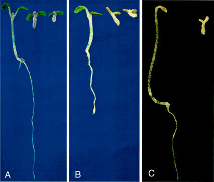
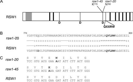
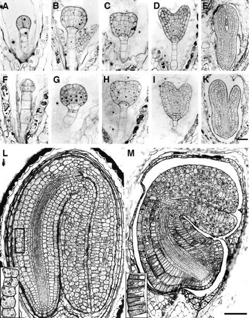
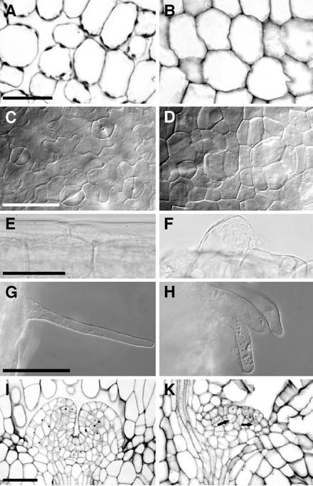
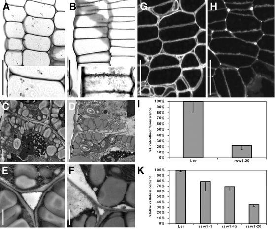
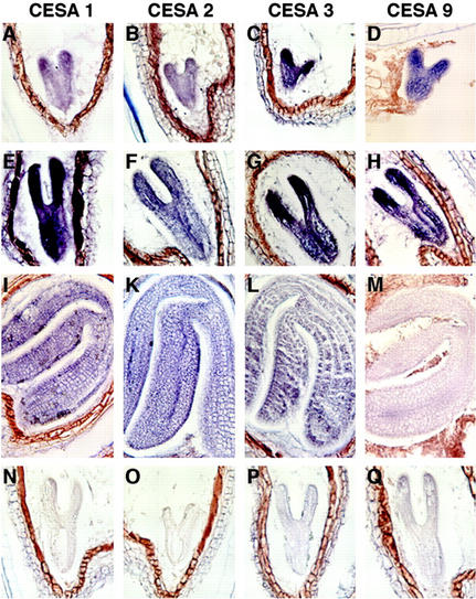
The products of the cellulose synthase A (CESA) gene family are thought to function as isoforms of the cellulose synthase catalytic subunit, but for most CESA genes, the exact role in plant growth is still unknown. Assessing the function of individual CESA genes will require the identification of the null-mutant phenotypes and of the gene expression profiles for each gene. Here, we report that only four of 10 CESA genes, CESA1, CESA2, CESA3, and CESA9 are significantly expressed in the Arabidopsis embryo. We further identified two new mutations in the RADIALLY SWOLLEN1 (RSW1/CESA1) gene of Arabidopsis that obstruct organized growth in both shoot and root and interfere with cell division and cell expansion already in embryogenesis. One mutation is expected to completely abolish the enzymatic activity of RSW1(CESA1) because it eliminated one of three conserved Asp residues, which are considered essential for beta-glycosyltransferase activity. In this presumed null mutant, primary cell walls are still being formed, but are thin, highly undulated, and frequently interrupted. From the heart-stage onward, cell elongation in the embryo axis is severely impaired, and cell width is disproportionally increased. In the embryo, CESA1, CESA2, CESA3, and CESA9 are expressed in largely overlapping domains and may act cooperatively in higher order complexes. The embryonic phenotype of the presumed rsw1 null mutant indicates that the RSW1(CESA1) product has a critical, nonredundant function, but is nevertheless not strictly required for primary cell wall formation.


Figures

Figure 1
Figure 1
Seedling phenotype of wild-type and rsw1 mutants 5 d after germination. A and B, Seedlings germinated in the light at 22°C (A) and 31°C (B). Genotypes from left to right are Ler wild type, rsw1-45, and rsw1-20. C, Ler wild-type and rsw1-20 mutant seedlings germinated at 22°C in the dark.
Figure 2
Figure 2
Mutations in the RSW1(CESA1) gene. A, Schematic diagram of RSW1(CESA1) protein indicating position and identity of the predicted altered amino acid residues in the rsw1-20 and rsw1-45 mutants. Gray, Conserved zinc finger; black, transmembrane domains. Bottom, Conserved amino acids in β-glycosyltranserases. B, Predicted amino acid sequence and secondary structure of rsw1-20 mutant and wild-type gene product. Bold, Conserved residues or motifs; dashed, sheet; dotted, coil; waved, helix. C, Nucleotide and amino acid sequence in mutant and wild-type alleles. Deviations from wild-type sequence are in bold, and amino acids are in one-letter code.
Figure 3
Figure 3
Wild-type (A–E and L) and rsw1-20 mutant (F–K and M) embryogenesis. A and F, Octant stage; defects in early embryos include abnormally oriented cell divisions. B through D and G through I, Globular and heart stages; largely normal cell arrangement, but mutant cells, particularly in the ground tissue of the hypocotyl, are generally somewhat shorter and radially expanded. E and K, Torpedo stage; pronounced length-to-width changes are evident in cells along the length of the embryo, particularly in the outer layers. L and M, Bent-cotyledon stage; short cotyledons with fewer cells along the length (for example, in the palisade mesophyll layer), which are, however, relatively normal in shape. In the hypocotyl and radicle, by contrast, the number of cells in the longitudinal dimension is nearly normal, whereas the length to width ratio of cells in nonvascular tissues is extremely distorted (compare wild-type and mutant cell number and cell shape in the outer cortex layer, enlarged in inlets). Arrow in inlet points at one of many incomplete cell divisions in the outer layers of the mutant hypocotyl. Scale bars = 20 μm in A, B, F, and G, same magnification; 50 μm in C, D, H, and I, same magnification; 75 μm in E and K; and 100 μm in M and L, same magnification.
Figure 4
Figure 4
Tissues in wild-type (A, C, E, G, and I) and rsw1-20 (B, D, F, H, and K) light-germinated seedlings 5 d after germination. A and B, Cross sections through cotyledon mesophyll. Intercellular spaces are strongly reduced, and no air cavities are formed in the mutant. C and D, Abaxial epidermis of cotyledons. Note the rounded cell shape and the absence of guard cells in the mutant. E and F, Hypocotyl epidermis cells, very regularly shaped in the wild type and generally irregular in the mutant. Within the irregular epidermal layer, some cells become extremely large. G and H, Root hairs are generally shorter and often thicker in the mutant. I and K, Shoot apical meristem, median section. Note fewer cells, incomplete cell divisions (arrows), and a highly irregular cell pattern in the mutant meristem. Except for a separation of the epidermal layer, no zonation is visible at this stage. Bright-field (A, B, E, F, I, and K) and whole-mount images with differential contrast optics (C, D, G, and H). Scale bars = 50 μm in A through H and 75 μm in I and K.
Figure 5
Figure 5
Cell wall structure and composition in wild-type and rsw1-20 mutant embryos. A and B, Periodic acid-Schiff's stained longitudinal sections in the hypocotyl of bent-cotyledon stage embryos. Note the presence of intercellular spaces in the wild type (A), which are absent in the mutant (B), and the uneven, granular appearance of mutant cell walls (inset). C thorough F, Epidermal cell walls from hypocotyls of bent-cotyledon stage embryos of wild type (C and E) and rsw1-20 (D and F). Note that mutant cell walls are thinner and that the middle lamella is usually not discernible. Intercellular spaces in the wild type (E) are usually filled with wall material in rsw1-20 mutants (F). G and H, Calcofluor stained cross sections in the hypocotyl of bent-cotyledon stage wild-type (G) and rsw1-20 mutant (H) embryos. Overall reduced staining in mutant walls indicates reduced β-glucan content. Note the uneven staining intensity in mutant cell walls. I, Densitometric quantification of cell wall β-glucan content measured on digital images from calcofluor-stained hypocotyl cells of bent-cotyledon stage embryos. K, Spectrophotometrical determination of β-glucan content in wall preparations of wild-type and mutant seedlings after germination and growth for 7 d at 31°C. A and B, Bright-field optics images; C through F, transmission electron micrographs; and G and H, epifluorescence optics. Scale bars = 20 μm in A, B, G, and H; insets, 2× magnification; 1 μm in C and D; and 500 nm in E and F.
Figure 6
Figure 6
CESA gene expression in embryos, young plants, and mature plants. Semiquantitative RT-PCR of total RNA prepared from embryos, seedlings, inflorescence stems, and flowers. Lanes 1 to 10, CESA1 to CESA10 RT-PCR products; R, approximately evenly expressed ROC1 gene (Lippuner et al., 1994); numbers on the right indicate the size of the PCR products in base pairs.
Figure 7
Figure 7
Expression pattern of CESA gene mRNA in wild-type (Col-0) embryos at heart/early torpedo (A–D), late torpedo (E–H), and bent-cotyledon (I–M) stage. Hybridization with antisense (A–M) and sense probes (N–Q). A, E, I, and N, RSW1(CESA1); B, F, K, and O, CESA2; C, G, L, and P, CESA3; and D, H, M, and Q, CESA9.

References

    1. Arioli T, Peng L, Betzner AS, Burn J, Wittke W, Herth W, Camilleri C, Höfte H, Plazinski J, Birch R et al. Molecular analysis of cellulose biosynthesis in Arabidopsis. Science. 1998;279:717–720. - PubMed
    1. Beeckman T, De Rycke R, Viane R, Inzé D. Histological study of seed coat development in Arabidopsis thaliana. J Plant Res. 2000;113:139–148.
    1. Burn JE, Hocart CH, Birch RJ, Cork AC, Williamson RE. Functional analysis of the cellulose synthase genes CesA1, CesA2, and CesA3 in Arabidopsis. Plant Physiol. 2002;129:797–807. - PMC - PubMed
    1. Carpita NC, Gibeaut DM. Structural models of primary cell walls in flowering plants: consistency of molecular structure with the physical properties of the walls during growth. Plant J. 1993;3:1–30. - PubMed
    1. Cutler S, Somerville C. Cellulose synthesis: cloning in silico. Curr Biol. 1997;7:R108–R111. - PubMed

Publication types

MeSH terms