. 2002 Dec;130(4):2188-98.
doi: 10.1104/pp.009654.

Biosynthesis of UDP-xylose. Cloning and characterization of a novel Arabidopsis gene family, UXS, encoding soluble and putative membrane-bound UDP-glucuronic acid decarboxylase isoforms

Affiliations

Biosynthesis of UDP-xylose. Cloning and characterization of a novel Arabidopsis gene family, UXS, encoding soluble and putative membrane-bound UDP-glucuronic acid decarboxylase isoforms

April D Harper et al. Plant Physiol. 2002 Dec.

Abstract

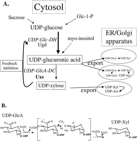
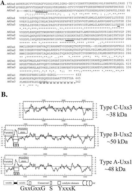
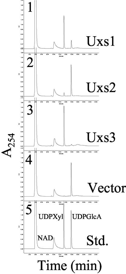
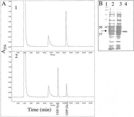
UDP-xylose (Xyl) is an important sugar donor for the synthesis of glycoproteins, polysaccharides, various metabolites, and oligosaccharides in animals, plants, fungi, and bacteria. UDP-Xyl also feedback inhibits upstream enzymes (UDP-glucose [Glc] dehydrogenase, UDP-Glc pyrophosphorylase, and UDP-GlcA decarboxylase) and is involved in its own synthesis and the synthesis of UDP-arabinose. In plants, biosynthesis of UDP-Xyl is catalyzed by different membrane-bound and soluble UDP-GlcA decarboxylase (UDP-GlcA-DC) isozymes, all of which convert UDP-GlcA to UDP-Xyl. Because synthesis of UDP-Xyl occurs both in the cytosol and in membranes, it is not known which source of UDP-Xyl the different Golgi-localized xylosyltransferases are utilizing. Here, we describe the identification of several distinct Arabidopsis genes (named AtUXS for UDP-Xyl synthase) that encode functional UDP-GlcA-DC isoforms. The Arabidopsis genome contains five UXS genes and their protein products can be subdivided into three isozyme classes (A-C), one soluble and two distinct putative membrane bound. AtUxs from each class, when expressed in Escherichia coli, generate active UDP-GlcA-DC that converts UDP-GlcA to UDP-Xyl. Members of this gene family have a large conserved C-terminal catalytic domain (approximately 300 amino acids long) and an N-terminal variable domain differing in sequence and size (30-120 amino acids long). Isoforms of class A and B appear to encode putative type II membrane proteins with their catalytic domains facing the lumen (like Golgi-glycosyltransferases) and their N-terminal variable domain facing the cytosol. Uxs class C is likely a cytosolic isoform. The characteristics of the plant Uxs support the hypothesis that unique UDP-GlcA-DCs with distinct subcellular localizations are required for specific xylosylation events.


Figures

Figure 1
Figure 1
A, Metabolic routes involved in the synthesis of UDP-Xyl. Synthesis of UDP-Xyl occurs both in the cytosol and in membrane-bound compartments. B, Possible intermediates (in brackets) involved in the enzymatic conversion of UDP-GlcA to UDP-Xyl, based on Feingold (1982).
Figure 2
Figure 2
Amino acid sequence comparison of AtUxs1 to AtUxs2 and AtUxs3. A, ClustalX alignment of the conserved amino acid sequences of AtUxs1, 2, and 3. The N terminus alignment between AtUxs1, 2, and 3 is not shown because of low amino acid sequence identity. Alignment of the AtUxs1 and 2 starts at amino acids 116 and 117, respectively; alignment of AtUxs1 and 3 starts at amino acids 120 and 30, respectively. Identical amino acid residues are indicated by an asterisk, and similar amino acid residues are indicated by a colon. The GxxGxxG and the Ser and YxxxK motifs are underlined. The dashed line demonstrates the unconserved C-terminal region. B, Comparison of the hydropathy plots of AtUxs 1, 2, and 3. Note the variable N-terminal region of AtUxs1 and 2 that spans from amino acid 1 to 48 and 1 to 43, respectively, followed by an approximately 16-amino acid hydrophobic region encoding the putative transmembrane domain (tm), which is followed by a variable “stem domain” in AtUxs1 and 2 that spans from amino acid 66 to 115 and 61 to 116.
Figure 3
Figure 3
AtUxs 1, 2, and 3 are UDP-GlcA-decarboxylase isozymes. Total soluble protein (10 μg) derived from E. coli expressing, separately: AtUXS1(Δ1–88), 1, Uxs1; AtUXS2(Δ1–95), 2, Uxs2; AtUXS3, 3, Uxs3; or control vector, 4, vector, was incubated for 60 min with 1 mm UDP-GlcA and 1 mm NAD+ for 60 min. The products of the reactions were separated over a Hypersil strong anion exchange (SAX)-HPLC column. 5, Elution positions of standard nucleotides and nucleotide sugars. The chromatography peak that migrated with the same retention time as authentic UDP-Xyl was collected and analyzed by 1H-NMR spectroscopy. Similar results were obtained using total soluble proteins or desalted protein fraction.
Figure 4
Figure 4
Purification of active UDP-GlcA-DC (AtUxs3). A, Purified AtUxs3 was incubated with 1 mm UDP-GlcA and 1 mm NAD+ for 0 (1) or 15 (2) min. The products of the reactions were separated over a Phenomenex SAX-HPLC column. The retention times of std UDP-Xyl and UDP-GlcA are indicated. B, SDS-PAGE of AtUxs3 during purification. Total soluble E. coli protein expressing AtUXS3 (lane 2) was desalted (lane 3) and purified over an Ni column (lane 4). Lane 1, Marker proteins with the indicated molecular masses. The molecular mass of recombinant AtUxs3 (42.6 kD; arrow) is larger than the native AtUxs3 (38.5 kD) because of the N-terminal His-6 amino acid tag fusion that facilitates purification.
Figure 5
Figure 5
Kinetic studies of AtUxs3. UDP-GlcA-DC assays were performed with increasing amounts of UDP-GlcA (0.125–4 mm). Duplicate assays were conducted for 15 min at 30°C. Lineweaver-Burk and Hanes-Woolf plots were obtained with an R2 > 0.96.
Figure 6
Figure 6
Effect of pH on the activity of AtUxs3. A, Assays were carried out for 20 min at 30°C with 1 mm UDP-GlcA in 0.1 m sodium-phosphate or Tris-HCl at the indicated pH values. B, Protein was pre-incubated with or without 0.5 mm NAD+ at the indicated pHs for 30 min on ice. The pH was then adjusted to neutrality, and 1 mm UDP-GlcA and 1 mm NAD+ were added. Duplicate reactions were incubated for 20 min at 30°C. The data are the mean and varied by no more than 5%. The relative amount of UDP-Xyl produced is plotted; 100% equals 41 nmol of UDP-Xyl produced.
Figure 7
Figure 7
Expression of AtUXS1, 2, and 3 in Arabidopsis. Total RNA isolated from flowers, stems, roots, and rosette leaves of fully mature plants (Leaf A, 6-week-old plants), or rosette leaves of 3-week-old plants (Leaf B) was used to amplify by RT-PCR AtUXS-specific cDNA. The following gene-specific transcripts were amplified by RT-PCR: 4, 1,317-bp AtUXS1; 3, 1,341-bp AtUXS2; and 2, 1,046-bp AtUXS3. As internal RT-PCR controls, the amplification of the CAB79762 gene whose cDNA sequence is expressed in all EST databases examined resulted in a predicted 1,302-bp DNA fragment (5), and a portion of the actin gene was RT-PCR amplified using degenerated primers (McKinney et al., 1995) to yield a 495-bp fragment (1). The data are representative of at least three independent RT-PCR reactions.

Similar articles

Cited by

References

    1. Altschul SF, Madden TL, Schaffer AA, Zhang J, Zhang Z, Miller W, Lipman DJ. Gapped BLAST and PSI-BLAST: a new generation of protein database search programs. Nucleic Acids Res. 1997;25:3389–3402. - PMC - PubMed
    1. Amino SI, Takeuchi Y, Kamamine A. Changes in enzyme activities involved in the formation and interconversion of UDP-sugars during cell cycle in a synchronous culture of Catharanthus roseus. Physiol Plant. 1985;64:111–117.
    1. Ankel H, Ankel E, Feingold DS, Schutzbach JS. Formation of UDP-d-xylose in algae. Biochim Biophys Acta. 1967;136:172–175. - PubMed
    1. Ankel H, Feingold DS. Biosynthesis of uridine diphosphate d-xylose: I. Uridine diphosphate glucuronate carboxy-lyase of wheat germ. Biochemistry. 1965;4:2468–2475. - PubMed
    1. Ankel H, Feingold DS. Biosynthesis of uridine diphosphate d-xylose: II. Uridine diphosphate d-glucuronate carboxy-lyase of Cryptococcus laurentii. Biochemistry. 1966;5:182–189. - PubMed

MeSH terms

Associated data