Comparative Study
. 2002 Jul;7(3):269-80.
doi: 10.1379/1466-1268(2002)007<0269:hspphb>2.0.co;2.

Heat shock protein-27 protects human bronchial epithelial cells against oxidative stress-mediated apoptosis: possible implication in asthma

Affiliations
Comparative Study

Heat shock protein-27 protects human bronchial epithelial cells against oxidative stress-mediated apoptosis: possible implication in asthma

Anna M Merendino et al. Cell Stress Chaperones. 2002 Jul.

Abstract

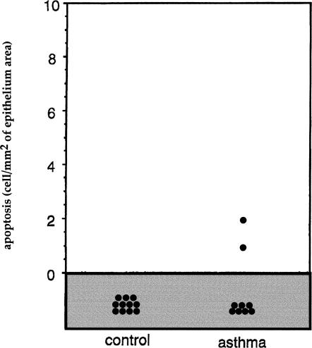
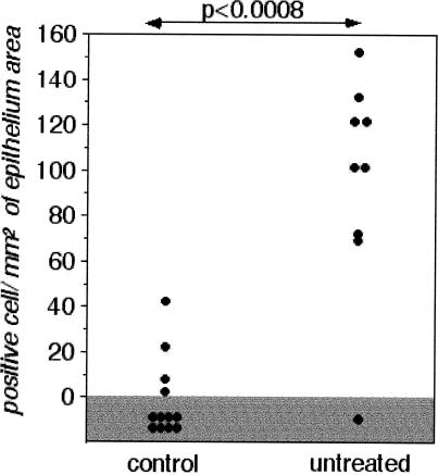
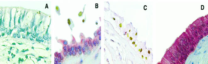
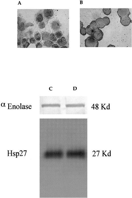
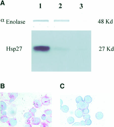
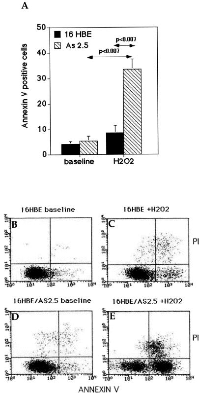
Inflammation of the human bronchial epithelium, as observed in asthmatics, is characterized by the selective death of the columnar epithelial cells, which desquamate from the basal cells. Tissue repair initiates from basal cells that resist inflammation. Here, we have evaluated the extent of apoptosis as well as the Hsp27 level of expression in epithelial cells from bronchial biopsy samples taken from normal and asthmatic subjects. Hsp27 is a chaperone whose expression protects against oxidative stress. We report that in asthmatic subjects the basal epithelium cells express a high level of Hsp27 but no apoptotic morphology. In contrast, apoptotic columnar cells are devoid of Hsp27 expression. Moreover, we observed a decreased resistance to hydrogen peroxide-induced apoptosis in human bronchial epithelial 16-HBE cells when they were genetically modified to express reduced levels of Hsp27.


Figures

Fig 1.
Fig 1.
Number of TUNEL-positive cells in bronchial biopsy specimens from control and asthmatic subjects. Sections of biopsy samples were prepared as described in Materials and Methods, and the number of TUNEL-positive epithelial cells was determined. Results are expressed as the number of TUNEL-positive cells per square millimeter of the epithelial area. Note the slight increase in the number of apoptotic cells in the biopsy samples from asthmatic patients. TUNEL, terminal deoxynucleotidyl transferase–mediated deoxynucleoside triphosphate nick end labeling
Fig 2.
Fig 2.
Number of Hsp27-positive cells. Expression of Hsp27 in epithelial cells of control and asthmatic subjects. Results are expressed as the number of Hsp27-positive cells per square millimeter of the epithelium. Statistical analysis by Mann-Whitney U-test
Fig 3.
Fig 3.
Analysis of Hsp27 expression and apoptosis in bronchial biopsy samples from normal and asthmatic patients. Hsp27 expression and TUNEL immunoreactivity in bronchial epithelial cells of a control (A) and an asthmatic subject (B, C, D) were performed as described in Materials and Methods. Cells expressing Hsp27 show a red immunostaining as the result of the labeled streptavidin-biotin immunoreactivity. TUNEL-positive cells are characterized by a brown staining of the nuclei (arrows). (A) Area of intact epithelium of a bronchial biopsy sample taken from normal subjects showing no immunoreactivity for Hsp27 and for TUNEL. (B) Area of damaged epithelium of a bronchial biopsy sample taken from asthmatic subjects with desquamated epithelial cells, which are not immunoreactive for Hsp27 but show a positivity for the TUNEL technique. (C) Wide area of fragile epithelium not immunoreactive for Hsp27 showing a nuclear TUNEL staining of many bronchial epithelial cells. (D) Area of intact epithelium showing a strong immunostaining for Hsp27, and a complete lack of nuclear staining by the TUNEL technique. TUNEL, terminal deoxynucleotidyl transferase–mediated deoxynucleoside triphosphate nick end labeling
Fig 4.
Fig 4.
Analysis of Hsp27 level in parental 16-HBE cells. Panels A and B: Immunocytochemical expression of Hsp27 before and after H2O2 (100 μM) exposure. Immunoreactivity, revealed by the labeled streptavidin biotin technique, is shown by the red staining of positive cells. Final magnification: 400×. Panels C and D: Western blot analysis of 16-HBE parental cells, 3 μg of each cellular extract was run on 10% SDS-PAGE and immunoblotted, as described in Materials and Methods. The relative intensity of the Hsp27 signal, compared with that of the housekeeping protein α-enolase, was of a factor of 100 before H2O2 exposure (panel C) and 110 after H2O2 (100 μM) exposure (panel D) in 16-HBE parental cells. 16-HBE, 16–human bronchial epithelial; SDS-PAGE, sodium dodecyl sulfate–polyacrylamide gel electrophoresis
Fig 5.
Fig 5.
Analysis of the level of Hsp27 expressed in the control and AS2.5 16-HBE cells. 16-HBE cells transfected with an empty vector are indicated as control cells. (A) Immunoblot analysis of alpha-enolase and Hsp27 cellular contents. Lane 1: 16-HBE control cells; lane 2: AS2.5 cells; lane 3: AS2.5 cells treated with H2O2 (100 μM for 1 hour and subsequently incubated for 18 hours without hydrogen peroxide). Note the sharp decline (about 80%) in the level of Hsp27 in AS2.5 cells and the disappearance of Hsp27 and enolase signals in H2O2-treated AS2.5 cells because of apoptosis-induced protein degradation. Immunocytochemical analysis of Hsp27 in AS2.5 cells (C) exposed or (B) not exposed to H2O2, as described earlier. Note the disappearance of the red staining after the hydrogen peroxide treatment. 16-HBE, 16–human bronchial epithelial
Fig 6.
Fig 6.
Analysis of the oxidative stress–mediated cell death of the control and AS2.5 16-HBE cells. (A) Estimation of the cell death of the control and Hsp27-underexpressing AS2.5 cells. Nontreated or H202-treated (100 μM for 1 hour, followed by 18 hours recovery) cells were resuspended in PBS-containing FITC-conjugated Annexin V and PI. Data are expressed as the percentage of Annexin V–positive–PI-negative cells. Black plot: control cells; hatched plot: AS2.5 cells. (B and C) Representative cytofluorimetric bivariate analysis of Annexin V binding and PI uptake in control cells at baseline (B) and after 100 μM H2O2 (1 hour, followed by 18 hours recovery) exposure (C). (D and E) As in (B) and (C), but in this case the analysis was performed using AS2.5 cells at baseline (D) and after 100 μM (1 hour) H2O2 treatment (E). 16-HBE, 16–human bronchial epithelial; PBS, phosphate-buffered saline; FITC, fluorescein isothiocyanate; PI, propidium iodide
Fig 7.
Fig 7.
Immunoblot analysis of caspase-3 activation. (a, b) Control or (c, d) AS2.5 cells were (b, d) exposed or (a, c) not exposed to 100 μM hydrogen peroxide during 1 hour, followed by 18 hours recovery. Cells were then harvested and total cellular proteins were applied to SDS-PAGE. Immunoblots were performed with the anti–caspase-3 antibody. The blots show that procaspase-3 is converted to its active form (17 kDa) in AS2.5 cells exposed to oxidative stress. A weak conversion is already detectable in nontreated AS2.5 cells. These phenomena were not detectable in control 16-HBE cells. 16-HBE, 16–human bronchial epithelial; SDS-PAGE, sodium dodecyl sulfate–polyacrylamide gel electrophoresis

References

    1. Arata S, Hamaguchi S, Nose K. Effects of the overexpression of the small heat shock protein, Hsp27, on the sensitivity of human fibroblast cells exposed to oxidative stress. J Cell Physiol. 1995;163:458–465. - PubMed
    1. Aron Y, Busson M, Polla BS, Dusser D, Lockhart A, Swierczewski E, Favatier F. Analysis of hsp70 gene polymorphism in allergic asthma. Allergy. 1999;54:165–170. - PubMed
    1. Arrigo A-P. Small stress proteins: chaperones that act as regulators of intracellular redox state and programmed cell death. Biol Chem. 1998;379:19–26. - PubMed
    1. Arrigo A-P. sHsp as novel regulators of programmed cell death and tumorigenicity. Pathol Biol. 2000;48:280–288. - PubMed
    1. Arrigo A-P. Small stress proteins: novel regulators of intracellular redox state. IUBMB Life. 2002;52:303–307. - PubMed

Publication types

MeSH terms