. 2002 Dec 15;545(3):837-53.
doi: 10.1113/jphysiol.2002.027235.

Regulation of Cl--HCO3- exchangers by cAMP-dependent protein kinase in adult rat hippocampal CA1 neurons

Affiliations

Regulation of Cl--HCO3- exchangers by cAMP-dependent protein kinase in adult rat hippocampal CA1 neurons

Christopher L Brett et al. J Physiol. .

Abstract

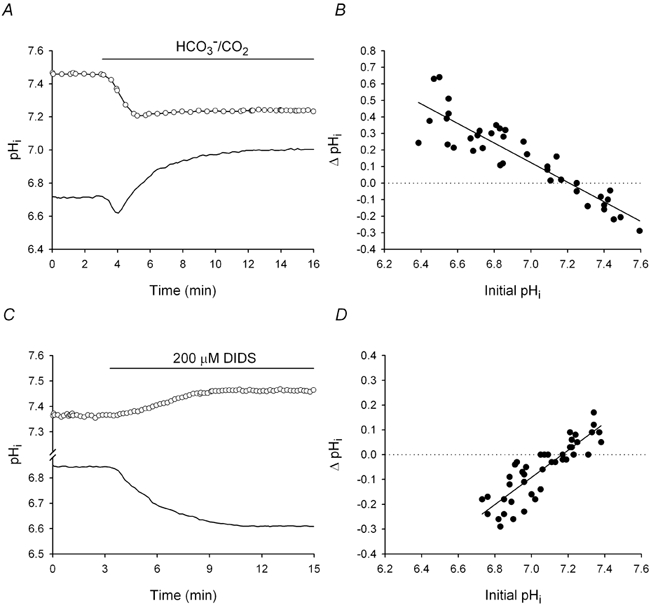
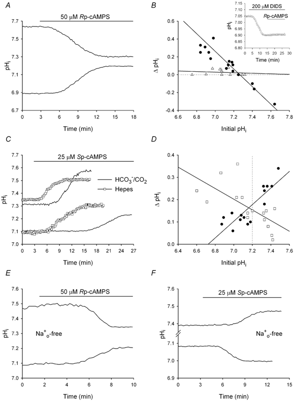
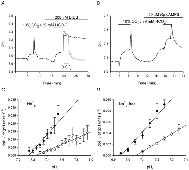
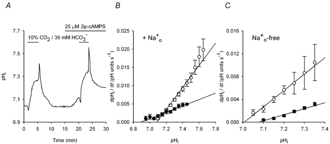
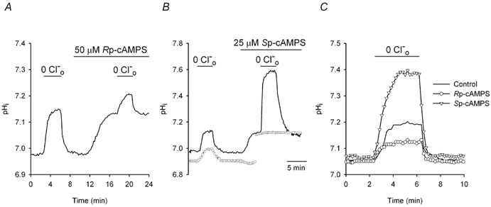
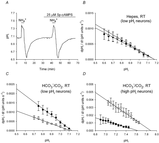
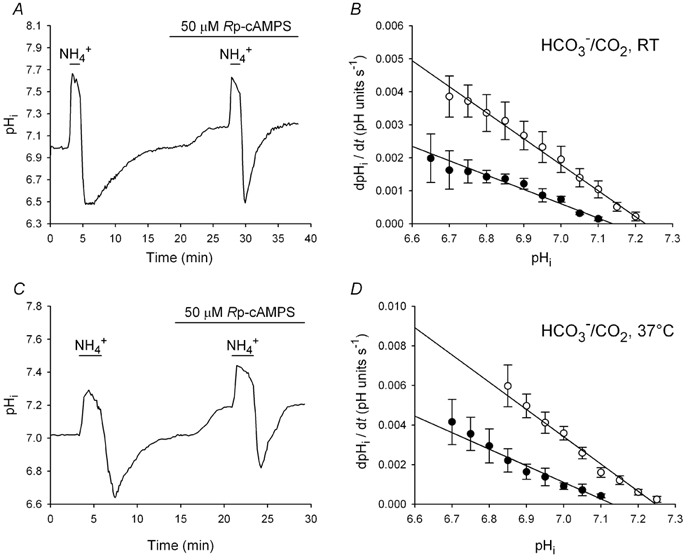
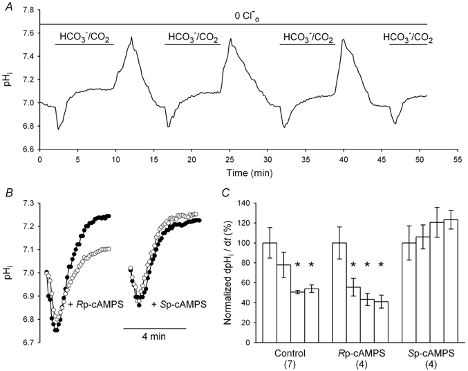
The contributions of HCO(3)(-)-dependent, DIDS-sensitive mechanisms to the maintenance of steady-state pH(i), and the regulation of their activities by cAMP-dependent protein kinase (PKA), were investigated in CA1 neurons with the H(+)-sensitive fluorophore, BCECF. The addition of HCO(3)(-)/CO(2) to neurons with "low" (pH(i) < or = 7.20) and "high" (pH(i) > 7.20) initial pH(i) values under Hepes-buffered conditions, increased and decreased steady-state pH(i), respectively. Conversely, under HCO(3)(-)/CO(2)-buffered conditions, DIDS caused pH(i) to decrease and increase in neurons with low and high initial pH(i) values, respectively. In the presence, but not the absence, of HCO(3)(-), the PKA inhibitor Rp-adenosine-3',5'-cyclic monophosphorothioate (Rp-cAMPS; 50 microM) evoked DIDS-sensitive increases and decreases in pH(i) in neurons with low and high initial pH(i) values, respectively. In contrast, in neurons with low initial pH(i) values, activation of PKA with the Sp isomer of cAMPS (Sp-cAMPS; 25 microM) elicited increases in pH(i) that were smaller in the presence than in the absence of HCO(3)(-), whereas in neurons with high initial pH(i) values, Sp-cAMPS-evoked rises in pH(i) were larger in the presence than in the absence of HCO(3)(-); the differences between the effects of Sp-cAMPS on pH(i) under the different buffering conditions were attenuated by DIDS. Consistent with the possibility that changes in the activities of HCO(3)(-)-dependent, DIDS-sensitive mechanisms contribute to the steady-state pH(i) changes evoked by the PKA modulators, in neurons with initial pH(i) values < or = 7.20, Rp-cAMPS concurrently inhibited Na(+)-independent Cl(-)-HCO(3)(-) exchange and stimulated Na(+)-dependent Cl(-)-HCO(3)(-) exchange; in contrast, Sp-cAMPS concurrently stimulated Na(+)-independent Cl(-)-HCO(3)(-) exchange and inhibited Na(+)-dependent Cl(-)-HCO(3)(-) exchange. Data from a limited number of neurons with initial pH(i) values > 7.20 suggested that the directions of the reciprocal changes in anion exchange activities (inhibition or stimulation) evoked by Rp- and Sp-cAMPS may be opposite in cells with low vs. high resting pH(i) values. Taken together, the results indicate that the effects of modulating PKA activity on steady-state pH(i) in rat CA1 neurons under HCO(3)(-)/CO(2)-buffered conditions reflect not only changes in Na(+)-H(+) exchange activity but also changes in Na(+)-dependent and Na(+)-independent Cl(-)-HCO(3)(-) exchange activity that, in turn, may be dependent upon the initial pH(i).


Figures

Figure 1
Figure 1. Effects on pHi of the transition from Hepes- to HCO3/CO2-buffered medium, and the addition of DIDS to HCO3/CO2-buffered medium
A, a neuron perfused with a Hepes-buffered medium (pH 7.35) had a low initial pHi (continuous line). Upon exposure to HCO3/CO2-buffered medium at a constant pHo, pHi decreased transiently and then increased to a new steady-state level. In contrast, a different neuron with a high initial pHi in Hepes-buffered medium (○) exhibited an internal acidification upon exposure to HCO3/CO2. B, the changes in pHi (ΔpHi) evoked by the addition of HCO3/CO2 plotted against initial pHi values in Hepes-buffered medium (n = 39). A linear least squares regression fit to the data points (r2= 0.84) had a negative slope and intersected the abscissa at pHi 7.21. C, in a neuron with a low initial pHi value under HCO3/CO2-buffered conditions at pHo 7.35 (continuous line), DIDS caused pHi to decrease to a new steady-state level. In contrast, DIDS caused pHi to rise in a different neuron with a high initial pHi (○). D, ΔpHi elicited by 200 μm DIDS applied under HCO3/CO2-buffered conditions plotted against initial pHi values (n = 44). A linear least squares regression fit to the data points (r2= 0.75) had a similar x-intercept (pHi 7.17) but an opposite slope to the fit representing the pHi response to HCO3/CO2 application.
Figure 2
Figure 2. Effects of Rp- and Sp-cAMPS on pHi
A, Rp-cAMPS, applied under HCO3/CO2-buffered conditions (pHo 7.35) for the period indicated by the bar above the traces, evoked increases and decreases in pHi in two different neurons with low and high initial pHi values, respectively, prior to the addition of the PKA inhibitor. B, the changes in pHi (ΔpHi) elicited by 50 μm Rp-cAMPS under HCO3/CO2-buffered control conditions (•) or following 10-15 min pre-treatment with 200 μm DIDS (▵) were plotted against pHi values measured prior to the addition of the PKA inhibitor and each set of data points was fitted by least squares regression (r2≥ 0.80). The changes in pHi evoked by Rp-cAMPS were dependent upon the initial pHi and, as illustrated in the inset, were attenuated by DIDS. C, four different neurons, two with similarly low initial pHi values and two with similarly high initial pHi values, were exposed to 25 μm Sp-cAMPS under HCO3/CO2- (continuous line) or Hepes- (□) buffered conditions. In neurons with initial pHi values ≤ 7.20, Sp-cAMPS evoked a greater increase in pHi under Hepes- than under HCO3/CO2-buffered conditions; in contrast, in neurons with initial pHi values > 7.20, Sp-cAMPS evoked a smaller increase in pHi under Hepes- than under HCO3/CO2-buffered conditions. D, ΔpHi elicited by 25 μm Sp-cAMPS in the nominal absence (□) or presence (•) of HCO3/CO2 were plotted against initial values of pHi and a regression line was fitted to each set of data points. The vertical dotted line represents the division between ‘low’ (initial pHi≤ 7.20) and ‘high’ (initial pHi > 7.20) pHi neurons. E, the same experiment as that shown in A, but conducted in the absence of external Na+. Rp-cAMPS evoked an increase and a decrease in pHi in two different neurons with low and high initial pHi values, respectively. F, under Na+o-free, HCO3-buffered conditions, the addition of 25 μm Sp-cAMPS caused pHi to increase in a neuron with a high initial pHi, and pHi to decrease in a different neuron with a low initial pHi.
Figure 3
Figure 3. Effects of Rp-cAMPS on pHi recovery from base loading in the presence and absence of Na+o
All data were obtained from neurons with initial pHi values ≤ 7.20 under HCO3/CO2-buffered conditions. Alkali loads were imposed by applying and withdrawing a 10 % CO2/39 mm HCO3 solution, as indicated by the short bars above the traces in A and B. A, following an initial alkali load, pHi was allowed to recover and a second alkali load was imposed in the presence of DIDS (applied for the period indicated by the long bar above the trace). Superimposed is a record (○) obtained from a different cell that was exposed to Cl-free medium (applied for the period indicated by the short bar beneath the trace) at the peak of the second alkali load (the first part of the record from this experiment has been omitted for clarity). B, after recovery from an initial alkali load imposed under control conditions, the neuron was exposed to 50 μm Rp-cAMPS. Rp-cAMPS evoked a rise in pHi (see Fig. 2A) and a second alkali load was then imposed; the rate of pHi recovery was decreased in the presence of Rp-cAMPS. C, mean rates of pHi recovery following alkali loads imposed in the absence (•) and presence (○) of 50 μm Rp-cAMPS plotted against absolute values of pHi. Data points were obtained from 14 experiments of the type illustrated in B; error bars represent s.e.m.D, mean rates of pHi recovery from alkali loads obtained in the absence (•) and presence (○) of 50 μm Rp-cAMPS under Na+o-free conditions plotted against absolute values of pHi. Data points were obtained from seven experiments of the type illustrated in B, except in the absence of external Na+; error bars represent s.e.m.
Figure 4
Figure 4. Effects of Sp-cAMPS on pHi recovery from base loading in the presence and absence of Na+o
All data were obtained from neurons with initial pHi values ≤ 7.20 under HCO3/CO2-buffered conditions. A, after the recovery of pHi from a control alkali load imposed under Na+o-containing conditions, 25 μm Sp-cAMPS evoked a slow increase in steady-state pHi (see Fig. 2C). A second alkali load was then imposed and pHi recovered at a faster rate, and to a new steady-state level, in the continued presence of Sp-cAMPS. B, the pHi dependence of pHi recovery under Na+o-containing conditions in the absence (•) and presence (○) of 25 μm Sp-cAMPS. Data points were obtained from nine experiments of the type illustrated in A; error bars represent s.e.m. The alkaline shift in the pHi-dependence of pHi recovery from alkali loads evoked by Sp-cAMPS reflects the increase in pHi elicited by Sp-cAMPS in the presence of Na+o in neurons with resting pHi values ≤ 7.20. C, mean rates of pHi recovery from base loading in the absence (•) and presence (○) of 25 μm Sp-cAMPS under Na+o-free conditions plotted against absolute values of pHi. Data points were obtained from five experiments of the type illustrated in A, except in the absence of external Na+. The acidic shift in the pHi-dependence of pHi recovery from alkali loads in the presence of Sp-cAMPS reflects the decrease in pHi elicited by Sp-cAMPS in the absence of Na+o in neurons with resting pHi values ≤ 7.20 (see Fig. 2F).
Figure 5
Figure 5. Effects of Rp- and Sp-cAMPS on changes in pHi evoked by removal of Clo
All data were obtained from neurons with initial pHi values ≤ 7.20 under HCO3/CO2-buffered conditions. A, the acute removal of Clo (for the period indicated by the short bar above the trace) evoked an increase in pHi which recovered upon the reintroduction of the anion. Rp-cAMPS (50 μm) was then applied and, after pHi increased to a new steady-state level (see Fig. 2A), the removal of Clo evoked an increase in pHi that was smaller than in the absence of the PKA inhibitor. Increases in pHi evoked by the acute removal of Clo in the presence of a PKA modulator were measured as the difference between the maximum pHi observed in the absence of Clo and the plateau pHi value observed following the reintroduction of Clo in the continued presence of the PKA modulator. B, a neuron with a low initial pHi (continuous line) was exposed to Cl-free medium, which caused an increase in pHi. Sp-cAMPS (25 μm) was then applied; the PKA activator evoked an increase in pHi (see Fig. 2C) and subsequent exposure to Cl-free medium caused a large internal alkalinization. Superimposed is a record (□) obtained from a different low pHi neuron in which 25 μm Sp-cAMPS was co-applied with 200 μm DIDS; the gap in the trace represents a 5.5 min break in the record. The rise in pHi observed during exposure to Cl-free medium in the presence of Sp-cAMPS was blocked by DIDS. C, the increases in pHi evoked by transient exposure to Cl-free medium are shown in three different neurons with similar initial pHi values under control conditions (continuous line) and following pre-treatment with 25 μm Sp-cAMPS (▿) or 50 μm Rp-cAMPS (○).
Figure 6
Figure 6. Effects of Sp-cAMPS on pHi recovery from acid loads at room temperature
A, two consecutive acid loads, the second in the presence of 25 μm Sp-cAMPS, were imposed on a neuron with a low initial pHi under Hepes-buffered conditions. Rates of pHi recovery from both acid loads were similar and, in contrast to the effects of Sp-cAMPS under Hepes-buffered conditions at 37 °C (see Smith et al. 1998), pHi failed to recover to a higher steady-state level in the presence of Sp-cAMPS. B, rates of pHi recovery from acid loads performed in the absence (•) and presence (○) of 25 μm Sp-cAMPS plotted against absolute values of pHi; data points were obtained from nine experiments of the type shown in A (error bars represent s.e.m.). Sp-cAMPS failed to significantly affect the rate of pHi recovery at any absolute value of pHi. C, the pHi dependence of pHi recovery under HCO3/CO2-buffered conditions at RT in the absence (•) and presence (○) of 25 μm Sp-cAMPS; data points were obtained from six experiments of the type shown in A, except in the presence of HCO3/CO2. Rates of pHi recovery were significantly reduced in the presence of Sp-cAMPS at all absolute values of pHi. D, rates of pHi recovery from acid loads imposed on high pHi neurons (n = 6) under HCO3/CO2-buffered conditions at RT in the absence (•) and presence (○) of 25 μm Sp-cAMPS plotted against absolute values of pHi. In contrast to its effect in low pHi neurons, Sp-cAMPS increased rates of pHi recovery in neurons with high initial pHi values.
Figure 7
Figure 7. Effects of Rp-cAMPS on pHi recovery from acid loads in low pHi neurons
A, an acid load was imposed on a low pHi neuron under HCO3/CO2-buffered conditions at RT and, after recovery of pHi, 50 μm Rp-cAMPS was applied. Rp-cAMPS increased both steady-state pHi and the rate of pHi recovery from the second acid load. B, the pHi dependence of the rate of pHi recovery from acid loads imposed under HCO3/CO2-buffered conditions at RT in the absence (•) and presence (○) of 50 μm Rp-cAMPS; data points were obtained from 12 experiments of the type shown in A. C, the same experiment as shown in A, but conducted at 37 °C. D, the pHi dependence of the rate of pHi recovery from acid loads performed at 37 °C in the absence (•) and presence (○) of 50 μm Rp-cAMPS; data points were obtained from eight experiments of the type shown in C. In contrast to observations made in the nominal absence of HCO3/CO2, Rp-cAMPS applied to low pHi neurons in the presence of HCO3/CO2 increased rates of pHi recovery at all absolute values of pHi and shifted the pHi dependence of the rate of pHi recovery in an alkaline direction.
Figure 8
Figure 8. Effects Rp- and Sp-cAMPS on alkalinizations evoked by HCO3/CO2 under Clo-free conditions
A, under Clo-free conditions, repeated exposures to HCO3/CO2 caused pHi to transiently decrease and then increase to a new steady-state level; the magnitude of the HCO3/CO2-evoked increase in pHi declined with each successive application, resulting in a progressive reduction in the rate at which pHi reached each new steady-state level (see C, Control). B, the effects on pHi of exposing two different low pHi neurons to HCO3/CO2 under Clo-free conditions in the presence of 50 μm Rp-cAMPS (left-hand side) or 25 μm Sp-cAMPS (right-hand side). Under each condition, filled circles identify the record obtained during the first exposure to HCO3/CO2; traces identified by open circles represent the changes in pHi observed during the second (Rp-cAMPS) or fourth (Sp-cAMPS) exposures to HCO3/CO2 in the respective series. C, rates of alkalinization observed during consecutive applications of HCO3/CO2 in the continuous absence of Clo were calculated at an absolute pHi value of 7.00 and normalized to the mean rate of alkalinization observed during the first application of HCO3/CO2 under each experimental condition. The resulting normalized rates of alkalinization are presented as percentage values (± s.e.m.) under control conditions and in the presence of 50 μm Rp-cAMPS and 25 μm Sp-cAMPS. (Number of neurons examined under each experimental condition). *P < 0.05 for the difference between the first normalized rate of alkalinization under a given experimental condition.

Similar articles

Cited by

References

    1. Alvarez BV, Fujinaga J, Casey JR. Molecular basis for angiotensin II-induced increase of chloride/bicarbonate exchange in the myocardium. Circulation Research. 2001;89:1246–1253. - PubMed
    1. Autere A-M, Lamsa K, Kaila K, Taira T. Synaptic activation of GABAA receptors induces neuronal uptake of Ca2+ in adult rat hippocampal slices. Journal of Neurophysiology. 1999;81:811–816. - PubMed
    1. Baxter KA, Church J. Characterization of acid extrusion mechanisms in cultured fetal rat hippocampal neurones. Journal of Physiology. 1996;493:457–470. - PMC - PubMed
    1. Bevensee MO, Cummins TR, Haddad GG, Boron WF, Boyarsky G. pH regulation in single CA1 neurons acutely isolated from the hippocampi of immature and mature rats. Journal of Physiology. 1996;494:315–328. - PMC - PubMed
    1. Boron WF, McCormick WC, Roos A. pH regulation in barNaCle muscle fibers: dependence on intracellular and extracellular pH. American Journal of Physiology. 1979;237:C185–193. - PubMed

Publication types

MeSH terms

LinkOut - more resources