. 2002 Dec 15;545(3):933-44.
doi: 10.1113/jphysiol.2002.029488.

Leptin inhibits epileptiform-like activity in rat hippocampal neurones via PI 3-kinase-driven activation of BK channels

Affiliations

Leptin inhibits epileptiform-like activity in rat hippocampal neurones via PI 3-kinase-driven activation of BK channels

L J Shanley et al. J Physiol. .

Abstract

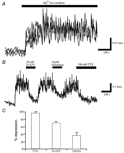
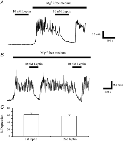
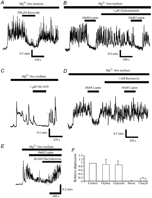
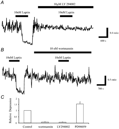
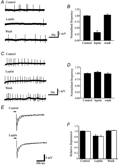
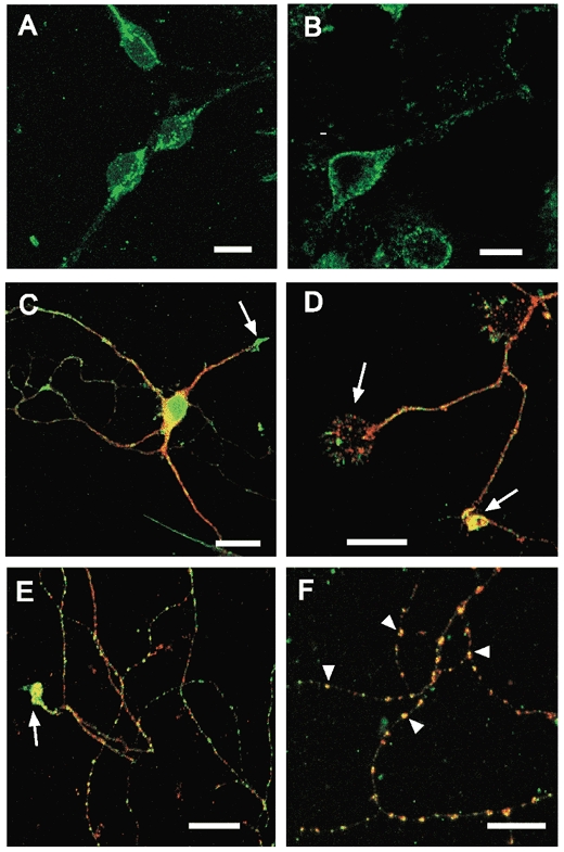
The obese gene product, leptin is an important circulating satiety factor that regulates energy balance via its actions in the hypothalamus. However, leptin receptors are also expressed in brain regions not directly associated with energy homeostasis, such as the hippocampus. Here, leptin inhibits hippocampal neurones via activation of large conductance Ca(2+)-activated K(+) (BK) channels, a process that may be important in regulating neuronal excitability. We now show that leptin receptor labelling is expressed on somata, dendrites and axons, and is also concentrated at synapses in hippocampal cultures. In functional studies, leptin potently and reversibly reduces epileptiform-like activity evoked in lean, but not leptin-resistant Zucker fa/fa rats. Furthermore, leptin also depresses enhanced Ca(2+) levels evoked following Mg(2+) removal in hippocampal cultures. The ability of leptin to modulate this activity requires activation of BK, but not K(ATP), channels as the effects of leptin were mimicked by the BK channel activator NS-1619, and inhibited by the BK channel inhibitors, iberiotoxin and charybdotoxin. The signalling mechanisms underlying this process involve stimulation of phosphoinositide 3-kinase (PI 3-kinase), but not mitogen-activated protein kinase (MAPK), as two structurally unrelated inhibitors of PI 3-kinase, LY294002 and wortmannin, blocked the actions of leptin. These data indicate that leptin, via PI 3-kinase-driven activation of BK channels, elicits a novel mechanism for controlling neuronal excitability. As uncontrolled excitability in the hippocampus is one underlying cause of temporal lobe epilepsy, this novel action of leptin could provide an alternative therapeutic target in the management of epilepsy.


Figures

Figure 1
Figure 1. Exposure of hippocampal cultures to Mg2+-free conditions induces spontaneous Ca2+ oscillations
A, perfusion of hippocampal cultures with Mg2+-free Hepes-buffered saline resulted in a rise in Ca2+ levels that was accompanied by the appearance of synchronised oscillations in Ca2+. Traces are derived from the somata of individual neurones within a field of cells. B, following generation of the spontaneous Ca2+ transients, application of TTX (500 nm) for the time indicated resulted in complete inhibition of the enhanced Ca2+ load associated with Mg2+ removal. Application of the NMDA receptor antagonist, D-APV (50 μm) or VGCC blocker, nifedipine (10 μm), attenuated the spontaneous activity in a reversible manner. C, pooled data illustrating the relative depressions of mean Ca2+ load in Mg2+-free medium following the addition of TTX (1 μm; n = 84), D-APV (50 μm; n = 80) or nifedipine (10 μm; n = 28). Thus, these synaptically driven Ca2+ transients required glutamatergic synaptic transmission as well as the influx of Ca2+ via VGCCs.
Figure 2
Figure 2. Leptin inhibits the enhanced Ca2+ levels evoked following Mg2+ removal
A, in control conditions (Mg2+-containing solution), application of leptin (10 nm) for the time indicated by the bar, had no effect the basal levels of Ca2+. However, following perfusion of Mg2+-free medium, which itself resulted in the generation of spontaneous Ca2+ oscillations, addition of leptin (10 nm) caused a rapid inhibition of the Ca2+ levels. This action of leptin was readily reversed on washout. B, the ability of leptin to inhibit this enhanced Ca2+ load was readily reproducible as subsequent application of leptin (10 nm), 20-30 min after the first exposure, resulted in a comparable response. C, pooled data illustrating that the relative depressions of mean Ca2+ load in Mg2+-free medium induced by sequential applications of leptin do not differ significantly.
Figure 3
Figure 3. Inhibition of Ca2+ levels by leptin involves BK, but not KATP, channel activation
A, following perfusion of Mg2+-free medium and the generation of the spontaneous Ca2+ oscillations, addition of the KATP channel opener, diazoxide (200 μm) reversibly depressed the mean enhancement of Ca2+ levels following Mg2+ removal. C, similarly, the BK channel activator, NS-1619 also depressed Ca2+ levels in a readily reversible manner. B and D, the KATP channel inhibitor, glybenclamide did not affect the leptin-modulation of Ca2+ levels, whereas the selective BK channel inhibitor, iberiotoxin completely blocked the actions of leptin. E, leptin-induced depression of the spontaneous Ca2+ transients was readily reversed by the addition of charybdotoxin (20 nm). F, pooled data illustrating the relative depressions of mean Ca2+ load in Mg2+-free medium by leptin in control conditions, and in the presence of glybenclamide (1 μm), glipizide (1 μm), iberiotoxin (1 nm) or charybdotoxin (20 nm).
Figure 4
Figure 4. Leptin inhibition of Ca2+ levels requires stimulation of PI 3-kinase
A, application of leptin (10 nm) for the time indicated caused reversible inhibition of the enhanced Ca2+ load in Mg2+-free medium. Exposure of the neurones to the PI 3-kinase inhibitor, LY 294002 (10 μm) for at least 15 min had no effect on the enhanced Ca2+ levels. However, subsequent addition of leptin in the continued presence of LY 294002 failed to inhibit Ca2+ levels. B, similarly, wortmannin (10 nm), another PI 3-kinase inhibitor had no effect on the enhanced Ca2+ levels, but it did significantly reduce the effects of leptin. C, pooled data illustrating the effects of leptin on the mean Ca2+ load in Mg2+-free medium in control conditions, and in the presence of wortmannin (10 nm; n = 67), LY 294002 (10 μm; n = 38) or the inhibitor of MAPK activation, PD 98059 (10 μm; n = 51).
Figure 5
Figure 5. Leptin inhibits interictal activity in Zucker lean, but not obese fa/fa rats
A, sample traces of extracellularly recorded spontaneous interictal events evoked in a hippocampal slice obtained from a Zucker lean rat. Leptin (50 nm) reduced the frequency of interictal activity in a readily reversible manner. B, plot of the pooled data illustrating the normalised interictal frequency in control conditions, in the presence of leptin (50 nm) and following washout (n = 4). C, sample traces of extracellularly recorded hippocampal epileptiform interictal activity in slices from obese Zucker fa/fa rats. Addition of leptin (50 nm) failed to affect the frequency of interictal events. D, pooled data illustrating the normalised frequency of interictal events in slices from fa/fa rats, in control conditions, in the presence of leptin and following washout (n = 4). E, sample records of evoked fEPSPs obtained in control conditions and following exposure to 50 nm leptin. Note that leptin not only reduced the slope and peak amplitude of the fEPSP, but it also reduced the number of population spikes. F, pooled data illustrating the relative depressions of the slope (▪) and amplitude (□) of fEPSPs evoked in control conditions, following exposure to leptin (50 nm) and on washout.
Figure 6
Figure 6. Leptin receptor localisation on cultured hippocampal neurones
A and B, leptin receptor immunoreactivity on neuronal somata, illustrating punctate labelling within the cytoplasm (A) and labelling associated with the plasma membrane (B). A is a Z-projection of a series of confocal images taken at 1 μm intervals, B is a single confocal section. Primary antibody labelling was visualised with an Alexa 488-conjugated donkey anti-goat secondary antibody (green). C, D, E and F, merged, single plane confocal images comparing leptin receptor labelling with monoclonal markers (red; Cy3) for soma/dendrites (MAP2; C), axons (GAP43, D) or synaptic terminals (synapsin 1; E and F). The yellow areas correspond to points of overlap. In these images, the confocal gain and/or laser intensity was increased to facilitate the visualisation of fine processes. Leptin receptor immunoreactivity was associated with synaptic markers in older cultures (9-14 days, E and F). In the zoomed image (F), arrowheads show examples of co-localisation with synapsin 1 at individual puncta. Strong leptin receptor immunoreactivity was also associated with dendritic (C) and axonal (D and E) growth cones (arrows). Scale bars are 10 μm (A, B, D, E and F) and 20 μm (C).

Similar articles

Cited by

References

    1. Abele AE, Miller RJ. Potassium channel activators abolish excitotoxicity in cultured hippocampal pyramidal neurons. Neuroscience Letters. 1990;115:195–200. - PubMed
    1. Abele AE, Scholz KP, Scholz WK, Miller RJ. Excitotoxicity induced by enhanced neurotransmission in cultured hippocampal pyramidal neurons. Neuron. 1990;4:413–419. - PubMed
    1. Ahima RS, Flier JS. Leptin. Annual Review Physiology. 2000;62:413–437. - PubMed
    1. Alessi DR, Cuendo A, Cohen P, Dudley DT, Saltiel AR. PD 98059 is a specific inhibitor of the activation of mitogen-activated protein kinase kinase in vitro and in vivo. Journal of Biological Chemistry. 1995;270:27489–27494. - PubMed
    1. Alger BE, Williamson AA. A transient calcium-dependent potassium component of the epileptiform burst after-hyperpolarisation in rat hippocampus. Journal of Physiology. 1988;399:191–205. - PMC - PubMed

Publication types

MeSH terms