. 2002 Dec 15;545(3):945-60.
doi: 10.1113/jphysiol.2002.023390.

Active and passive membrane properties of rat sympathetic preganglionic neurones innervating the adrenal medulla

Affiliations

Active and passive membrane properties of rat sympathetic preganglionic neurones innervating the adrenal medulla

Jennifer M M Wilson et al. J Physiol. .

Abstract

The intravascular release of adrenal catecholamines is a fundamental homeostatic process mediated via thoracolumbar spinal sympathetic preganglionic neurones (AD-SPN). To understand mechanisms regulating their excitability, whole-cell patch-clamp recordings were obtained from 54 retrogradely labelled neonatal rat AD-SPN. Passive membrane properties included a mean resting membrane potential, input resistance and time constant of -62 +/- 6 mV, 410 +/- 241 MOmega and 104 +/- 53 ms, respectively. AD-SPN were homogeneous with respect to their active membrane properties. These active conductances included transient outward rectification, observed as a delayed return to rest at the offset of the membrane response to hyperpolarising current pulses, with two components: a fast 4-AP-sensitive component (A-type conductance), contributing to the after-hyperpolarisation (AHP) and spike repolarisation; a slower prolonged Ba(2+)-sensitive component (D-like conductance). All AD-SPN expressed a Ba(2+)-sensitive instantaneous inwardly rectifying conductance activated at membrane potentials more negative than around -80 mV. A potassium-mediated, voltage-dependent sustained outward rectification activated at membrane potentials between -35 and -15 mV featured an atypical pharmacology with a component blocked by quinine, reduced by low extracellular pH and arachidonic acid, but lacking sensitivity to Ba(2+), TEA and intracellular Cs(+). This quinine-sensitive outward rectification contributes to spike repolarisation. Following block of potassium conductances by Cs(+) loading, AD-SPN revealed the capability for autorhythmicity and burst firing, mediated by a T-type Ca(2+) conductance. These data suggest the output capability is dynamic and diverse, and that the range of intrinsic membrane conductances expressed endow AD-SPN with the ability to generate differential and complex patterns of activity. The diversity of intrinsic membrane properties expressed by AD-SPN may be key determinants of neurotransmitter release from SPN innervating the adrenal medulla. However, factors other than active membrane conductances of AD-SPN must ultimately regulate the differential ratio of noradrenaline (NA) versus adrenaline (A) release secreted in response to various physiological and environmental demands.


Figures

Figure 1
Figure 1. Intracellular and retrograde labels confirm recorded SPN innervate the adrenal medulla
A, longitudinal section of thoracolumbar spinal cord (35 μm thick) showing the distribution of SPN innervating the adrenal medulla as revealed by retrograde labelling of SPN with Rhodamine Dextran Lysine (RDL). All labelled SPN were observed in the spinal cord ipsilateral to the injected gland. B, transverse section (25 μm) of thoracic spinal cord showing SPNs labelled with RDL after injection into the adrenal medulla. The neurone indicated with the arrow in a was also filled (b) with Lucifer Yellow (LY) from the recording pipette, identifying its axonal projection to the adrenal medulla. In the latter section, note another LY-labelled neurone that lacked RDL, therefore not considered among the AD-SPN population data. Note, one neurone per slice was usually recorded except in instances to confirm the lack of overlap between the dyes used for retrograde and intracellular labelling. Scale bars are 50 μm in A and 25 μm B.
Figure 2
Figure 2. Membrane properties characteristic of AD-SPN
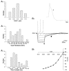
A, frequency histograms summarising the distribution of passive membrane properties of AD-SPN. a, resting membrane potentials (RMP; mV). b, input resistances (MΩ). c, membrane time constants (ms). B, whole-cell current-clamp recordings (holding potential -50 mV) illustrate characteristic active membrane properties. a, instantaneous inward rectification (*) activated during the membrane response to large amplitude hyperpolarising current pulses (not shown), and transient outward rectification (▪) observed as a delayed return to rest of the membrane responses. Also shown in the inset is an action potential on a faster time base to illustrate the pronounced shoulder on the repolarising phase (+). b, a steady-state I-V curve plotted from data recorded at the end of membrane responses to current injection shown above reveals the inward rectification activated around -80 mV.
Figure 3
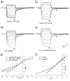
Figure 3. Instantaneous inward rectification in AD-SPN is mediated by a Ba2+- and Cs+-sensitive K+ conductance
A, samples of voltage traces recorded in the presence of TTX (0.5 μm) illustrate membrane responses to injection of current pulses (-200 to 100 pA, 20 pA increments) from a holding potential of -45 mV. Note in a instantaneous inward rectification activated in response to large amplitude current pulses and in b its reduction in the presence of extracellular barium. B, steady-state I-V curves illustrate pronounced inward rectification in control (□) and its subsequent blockade in the presence of Ba2+ (▵). Also note the absence of instantaneous inward rectification shown in an I-V curve for Cs+-loaded neurones (n = 12, •). C, voltage -current relations of an AD-SPN in the presence of 3.1 mm extracellular K+ (a) and nominally zero K+-containing ACSF (b). Membrane responses were evoked by injection of rectangular-wave current pulses (-120 to +100 pA, 20 pA increments, not shown) from a holding potential of -50 mV in the presence of TTX (0.5 μm). Note the enhanced transient outward rectification (▪) and reduced instantaneous inward rectification (*) following the manipulation of the K+ ion concentration gradient. The data plotted in c for the above illustrate instantaneous inward rectification (□) and its reduction in nominally zero K+-containing bathing medium (▴).
Figure 4
Figure 4. Sensitivity of the transient outward rectification to extracellular Ba2+ and 4-AP
A, voltage traces illustrate the transient outward rectification as a delayed return to the resting or holding potential at the break of the membrane response to hyperpolarising current pulses (20 pA steps) to the holding membrane potential (-50 mV; a); and its partial reduction in the presence of Ba2+. Subsequent addition of 4-AP (c) reduced further the remaining transient outward rectification. B, superimposed samples of membrane responses to hyperpolarising current pulses reveal the sensitivity of a slow component of the transient outward rectification to Ba2+ and a relatively fast component sensitive to subsequent addition of 4-AP (a). Traces in b illustrate the resistance of the slow component to 4-AP.
Figure 5
Figure 5. The transient outward rectification is insensitive to intracellular Cs+
A, voltage-current relations of a SPN in which intracellular K+ was replaced with equimolar Cs+ (140 mm; a) and subsequently after reducing extracellular K+ from 3.1 mm to nominally zero K+-containing ACSF (b). Membrane responses were evoked by injection of rectangular-wave current pulses (-220 to +280 pA, 20 pA increments) in the presence of TTX (1.0 μm). Note the shift in polarity of the transient outward rectification in the presence of intracellular Cs+ and its subsequent reversal in nominally zero K+-containing ACSF (arrows). B, superimposed membrane responses to depolarising current pulses (20 pA steps) following removal of inactivation by hyperpolarising current injection (-200 pA) to membrane potentials around -100 mV in Cs+-loaded SPN. In a note the transient depolarisation activated around -65 mV, delayed depolarisation around -45 mV and rebound depolarisation at the break of the response to the hyperpolarising step. In b, subsequent exposure of the slice to nominally 0 mm K+-containing ACSF recovered a transient outward rectification activated at membrane potentials around -65 mV and at the offset of the membrane response to the hyperpolarising current injection.
Figure 6
Figure 6. Two distinct components mediate the rebound depolarization revealed in Cs+-loaded AD-SPN
A, superimposed membrane responses to depolarising current pulses (20 pA steps) following removal of inactivation by hyperpolarising current injection (-200 pA) to membrane potentials around -100 mV in Cs+-loaded SPN. In a note the depolarisation activated around -65 mV and rebound depolarisation at the break of the response to the hyperpolarising step. * In b, subsequent application of 4-AP (4 mm) revealed a smaller more transient depolarisation activated around -55 to -60 mV and transient rebound depolarisation at the break of the response to membrane hyperpolarisation which could give rise to firing *. B, superimposed records showing the rebound depolarisations evoked at the offset of the membrane response to hyperpolarisation. Note the transient nature of the rebound depolarisation in the presence of 4-AP compared with control.
Figure 7
Figure 7. Burst firing induced in Cs+-loaded AD-SPN is mediated by a Ni2+-sensitive low-threshold Ca2+ conductance
A, spontaneous burst firing pattern of activity in Cs+-loaded AD-SPN. B, superimposed membrane responses to depolarising current pulses (20 pA steps) following removal of inactivation by hyperpolarising current injection (-100 pA) to membrane potentials around -100 mV in Cs+-loaded AD-SPN. In a, note the low-threshold depolarisation activated around -50 mV and rebound depolarisation at the break of the response to the hyperpolarising step. In b, note the transient depolarisation was completely blocked by subsequent exposure to Ni2+. C, spontaneous activity in Cs+-loaded AD-SPN (a) was completely (b) reversibly (c) blocked by Ni2+.
Figure 8
Figure 8. A sustained voltage-dependent outward rectifying K+ conductance in AD-SPN
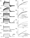
A, samples of a continuous whole-cell current-clamp recording illustrate superimposed membrane responses to injection of current pulses in the presence of 4-AP, TEA, Co2+ and TTX (1 μm; a). Note the persistent outward rectification in response to depolarising current pulses observed under these recording conditions. In the plot of these data (b), note the decrease in slope conductance at membrane potentials less negative than around -40 mV. B, superimposed membrane responses showing voltage-current relations of a AD-SPN in the presence of 3.1 mm extracellular K+ (a) and subsequently in elevated extracellular K+ (15 mm; (b)). Membrane responses were evoked by injection of depolarising current pulses (10 pA steps in a, 2 pA steps in b) in the presence of TTX (1 μm), TEA (30 mm), Ni2+ (2 mm) and 4-AP (4 mm). Note the strong outward rectification in control was reduced following exposure to elevated extracellular K+. C, plot of data shown in B and from data recorded in the same cell in reduced extracellular K+ (0.6 mm).
Figure 9
Figure 9. The effects of ion channel blockers on the sustained outward rectification
A, effects of ion channel blockers and ion substitutions on outward rectification evoked in response to membrane depolarisation from holding potentials around -50 mV. Superimposed membrane responses to depolarising current pulses (10 pA steps, a), revealed a transient early outward rectification leading to a delay to the peak of the membrane response, and a sustained rectification at the peak of the membrane response. In the presence of Co2+, the transient rectification was more obvious (b), whereas its subsequently blockade by 4-AP (c) left a sustained outward rectification. In d, high concentrations of TEA partly reduced the outward rectification, but sustained outward rectification remained. B, plots of data shown in A measured at the peak of the membrane response in control and at the same time point for subsequent manipulations (*), and during the steady-state membrane response (+). C, sustained outward rectification persisted when intracellular K+ was exchanged for equimolar Cs+ (a), in nominally 0 mm extracellular Ca2+ and reduced extracellular Na+, but was completely blocked by quinine (b). Da, pooled data from I-V plots showing the effects of intracellular Cs+ on outward rectification compared with control recording conditions with K+-gluconate-based pipette solution (n = 12); b, pooled data illustrate the effects of quinine on sustained outward rectification (n = 4).
Figure 10
Figure 10. The effects of changes in extracellular pH and arachidonic acid on the sustained outward rectification
A, samples of a continuous whole-cell current-clamp record, in the presence of TTX (0.5 μm), showing membrane responses to injection of current pulses (10 pA increments) from a holding potential of -50 mV. Membrane depolarisation evoked a sustained outward rectification (a) at pH 7.4. Reducing extracellular pH to 6.2 increased neuronal input resistance at potentials less negative than around -40 mV. At potentials less negative than around -25 mV a sustained outward rectification persisted (b). c, plot of data shown above: extracellular pH 7.4 (□) and pH 6.2 (▴). B, sustained outward rectification was sensitive to arachidonic acid. Membrane depolarisation evoked a sustained outward rectification (a)that was reduced in the presence of arachidonic acid (100 μm) (b). Membrane responses were evoked by depolarizing current pulses (10 pA steps) from holding potentials of -40 mV, in the presence of TTX (1 μm), Ni2+ (2 mm) and 4-AP (4 mm) to block voltage-dependent sodium, low-threshold calcium and transient outward rectification respectively. C, plot of pooled data from three neurones to illustrate the effects of arachidonic acid on the outward rectification: control (□); arachidonic acid (▴).
Figure 11
Figure 11. The role of outwardly rectifying conductances in action potential repolarisation and afterhyperpolarisation
Samples of a continuous whole-cell current-clamp recording illustrate the effects of ion channel blockers on the action potential and after-hyperpolarisation (AHP) waveforms. Each record is the average of 10 antidromic action potentials evoked by stimulation (9 V, 5 ms, 0.05 Hz) of the ventral horn exit zone. All records were obtained in the presence of D-AP5 (10 μm), NBQX (5 μm), bicuculline (10 μm) and strychnine (2 μm) to block fast chemical synaptic transmission. A, the evoked antidromic spike (1) was prolonged in the presence of 30 mm TEA (2) and further prolonged in the presence of both TEA and 4 mm 4-AP (3). In b, the same data as above but shown on a slower time base to illustrate the reduction in the AHP in the presence of TEA and 4-AP (3). B, recordings from another AD-SPN showing the effects of quinine on spike repolarisation. The evoked antidromic spike (1) was prolonged in the presence of 2 mm Ba2+ and 4 mm 4-AP (2) and further prolonged when 100 μm quinine was added in addition (3).

Similar articles

Cited by

References

    1. An WF, Bowlby MR, Betty M, Cao J, Ling HP, Mendoza G, Hinson JW, Mattsson KI, Strassle BW, Trimmer JS, Rhodes KJ. Modulation of A-type potassium channels by a family of calcium sensors. Nature. 2000;403:553–556. - PubMed
    1. Backman SB, Sequeira-Martinho H, Henry JL. Adrenal versus nonadrenal sympathetic preganglionic neurones in the lower thoracic intermediolateral nucleus of the cat: physiological properties. Canadian Journal of Physiology and Pharmacology. 1990;68:1447–1456. - PubMed
    1. Bahring R, Dannenberg J, Peters HC, Leicher T, Pongs O, Isbrandt D. Conserved Kv4 N-terminal domain critical for effects of Kv channel-interacting protein 2. 2 on channel expression and gating. Journal of Biological Chemistry. 2001;276:23 888–23 894. - PubMed
    1. Bekkers JM. Distribution and activation of voltage-gated potassium channels in cell-attached and outside-out patches from large layer 5 cortical pyramidal neurones of the rat. Journal of Physiology. 2000;525:611–620. - PMC - PubMed
    1. Block BM, Jones SW. Delayed rectifier current of bullfrog sympathetic neurones: ion-ion competition, asymmetrical block and effects of ions on gating. Journal of Physiology. 1997;499:403–416. - PMC - PubMed

Publication types

MeSH terms

LinkOut - more resources