. 2002 Dec 15;545(3):975-86.
doi: 10.1113/jphysiol.2002.031823.

Smooth muscle membrane potential modulates endothelium-dependent relaxation of rat basilar artery via myo-endothelial gap junctions

Affiliations

Smooth muscle membrane potential modulates endothelium-dependent relaxation of rat basilar artery via myo-endothelial gap junctions

Tracy Allen et al. J Physiol. .

Abstract

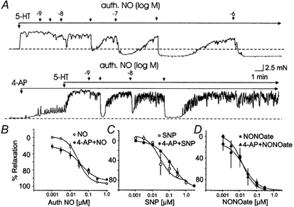
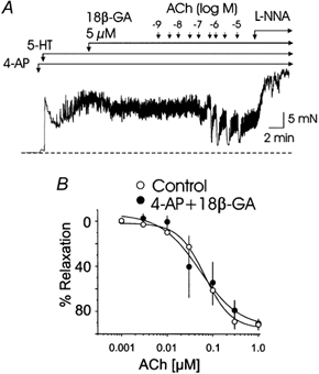
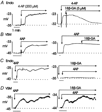
The release of endothelium-derived relaxing factors, such as nitric oxide (NO), is dependent on an increase in intracellular calcium levels ([Ca(2+)](i)) within endothelial cells. Endothelial cell membrane potential plays a critical role in the regulation of [Ca(2+)](i) in that calcium influx from the extracellular space is dependent on membrane hyperpolarization. In this study, the effect of inhibition of vascular smooth muscle delayed rectifier K(+) (K(DR)) channels by 4-aminopyridine (4-AP) on endothelium-dependent relaxation of rat basilar artery to acetylcholine (ACh) was assessed. ACh-evoked endothelium-dependent relaxations were inhibited by N-(Omega)-nitro-L-arginine (L-NNA) or 1H-[1,2,4]oxadiazolo[4,3-a]quinoxalin-1-one (ODQ), confirming a role for NO and guanylyl cyclase. 4-AP (300 microM) also suppressed ACh-induced relaxation, with the maximal response reduced from approximately 92 to approximately 33 % (n = 11; P < 0.01). However, relaxations in response to exogenous NO, applied in the form of authentic NO, sodium nitroprusside or diethylamineNONOate (DEANONOate), were not affected by 4-AP treatment (n = 3-11). These data are not consistent with the view that 4-AP-sensitive K(DR) channels are mediators of vascular hyperpolarization and relaxation in response to endothelium-derived NO. Inhibition of ACh-evoked relaxation by 4-AP was reversed by pinacidil (0.5-1 microM; n = 5) or 18beta-glycyrrhetinic acid (18betaGA; 5 microM; n = 5), indicating that depolarization and electrical coupling of the smooth muscle to the endothelium were involved. 4-AP caused depolarization of both endothelial and vascular smooth muscle cells of isolated segments of basilar artery (mean change 11 +/- 1 and 9 +/- 2 mV, respectively; n = 15). Significantly, 18betaGA almost completely prevented the depolarization of endothelial cells (n = 6), but not smooth muscle cells (n = 6) by 4-AP. ACh-induced hyperpolarization of endothelium and smooth muscle cells was also reduced by 4-AP, but this inhibition was not observed in the combined presence of 4-AP and 18betaGA. These data indicate that 4-AP can induce an indirect inhibition of endothelium-dependent relaxation in the rat basilar artery by electrical coupling of smooth muscle membrane depolarization to the endothelium via myo-endothelial gap junctions.


Figures

Figure 1
Figure 1. Effect of l-NNA (100 μm) and ODQ (10 μm) on ACh-evoked relaxation of segments of rat basilar artery precontracted with 5-HT
A, representative recordings showing ACh (1 nm to 3 μm)-evoked relaxations of 5-HT (0.1 μm)-induced tone in the absence and presence of l-NNA. B and C, cumulative concentration-relaxation curves for ACh in the absence and presence of l-NNA and l-NNA +l-NAME (100 μm), and ODQ, respectively. Data are the means of 14, 4 and 13 experiments, respectively, ± s.e.m. (in some cases the error bars in this and subsequent figures are within the size of the data symbols).
Figure 2
Figure 2. Effect of 4-AP (300 μm) on ACh-evoked relaxation of segments of rat basilar artery precontracted with 5-HT
A, representative recording showing oscillatory increase in basal tension induced by 4-AP in some tissues. B, representative recording showing ACh (1 nm to 3 μm)-evoked relaxations of 5-HT (0.1 μm)-induced tone in the absence and presence of 4-AP. C, cumulative concentration-relaxation curves for ACh in the absence and presence of 4-AP and clofilium (3 μm). Data are the average of 11 and 5 experiments ± s.e.m. with 4-AP and clofilium, respectively,
Figure 3
Figure 3. Effect of 4-AP (300 μm) on relaxations in response to NO in rat basilar artery
A, representative recordings showing authentic NO (1 nm to 1 μm)-induced relaxations of 5-HT (0.03 μm)-induced tone in the absence and presence of 4-AP. B, C and D, mean cumulative concentration-relaxation curves for authentic NO, SNP and DEANONOate (NONOate) in the absence and presence of 4-AP. Each point is the mean of 11, 5 and 3 experiments, respectively, ±s.e.m.
Figure 4
Figure 4. Effect of pinacidil (1 μm) on 4-AP-induced inhibition of relaxation in response to ACh in rat basilar artery
A, representative recordings showing the prevention of 4-AP (300 μm)-induced inhibition of ACh (1 nm to 10 μm)-evoked relaxation of 5-HT (0.3 μm)-induced tone by pinacidil (1 μm). Note that relaxation of 4-AP-induced increase in basal tone was unaffected by 0.1, but it was reversed by 1 μm pinacidil. B, mean cumulative concentration-relaxation curves for ACh in the absence and presence of 4-AP and pinacidil (1 μm). Each point is the mean of 5 experiments ±s.e.m.
Figure 5
Figure 5. Effect of 18βGA (5 μm) on 4-AP-induced inhibition of relaxation in response to ACh in rat basilar artery
A, representative recordings showing the prevention of 4-AP (300 μm)-induced inhibition of ACh (1 nm to 10 μm)-evoked relaxations of 5-HT (0.3 μm)-induced tone by 18βGA (5 μm). B, mean cumulative concentration-relaxation curves for ACh in the absence and presence of 4-AP and 18βGA. Each point is the mean of 5 experiments ±s.e.m.
Figure 6
Figure 6. Effect of BayK8644 (1 μm) on membrane potential of endothelial and smooth muscle cells of rat basilar artery in the absence and presence of 18βGA (5 μm)
Representative microelectrode recordings of BayK8644-induced depolarization of endothelial (Endo) and smooth muscle cells (VSM) in the absence (left) and presence (right) of 18βGA. Note that the depolarization of the endothelial cell, but not the VSM cell, due to BayK8644 was prevented by 18βGA.
Figure 7
Figure 7. Effect of 4-AP (200 μm) on membrane potential of endothelial and smooth muscle cells of rat basilar artery in the absence and presence of 18βGA (5 μm)
A, representative microelectrode recordings showing 4-AP-induced depolarization of an endothelial cell (left) and the ability of 18βGA to reverse the depolarization caused by 4-AP. B, representative microelectrode recordings showing 4-AP-induced depolarization of a vascular smooth muscle cell (left) and the inability of 18βGA to reverse the depolarization caused by 4-AP. C, representative microelectrode recordings showing 4-AP-induced depolarization of an endothelial cell (left) and the lack of depolarization in response to 4-AP in the presence of 18βGA. D, representative microelectrode recordings showing 4-AP-induced depolarization of a vascular smooth muscle cell in the absence (left) and presence of 18βGA (right).
Figure 8
Figure 8. Effect of l-NNA (100 μm) and 4-AP (200 μm) on ACh-induced hyperpolarization of the membrane potential of endothelial and smooth muscle cells of rat basilar artery
A, representative microelectrode recordings showing an ACh (bolus concentration 100 μmol)-induced hyperpolarization of a smooth muscle cell (VSM) in the absence (left) and lack of change in membrane potential following ACh application in the presence of l-NNA (right). B, representative microelectrode recordings of ACh-induced hyperpolarizations of an endothelial cell (Endo) in the absence (left) and in the presence of 4-AP alone (middle) and 4-AP + 18βGA (right). C, representative microelectrode recordings of ACh-induced hyperpolarization of a smooth muscle cell (VSM) in the absence (left) and in the presence of 4-AP (midde) and 4-AP + 18βGA (right).

References

    1. Adams DJ, Barakeh J, Laskey R, Van Breeman C. Ion channels and regulation of intracellular calcium in vascular endothelial cells. FASEB Journal. 1989;3:2389–2400. - PubMed
    1. Aiello EA, Malcolm AT, Walsh MP, Cole WC. Beta-adrenoceptor activation and PKA regulate delayed rectifier K+ channels of vascular smooth muscle cells. American Journal of Physiology. 1998;275:H448–459. - PubMed
    1. Aiello EA, Walsh MP, Cole WC. Phosphorylation by protein kinase A enhances delayed rectifier K+ current in vascular smooth muscle cells. American Journal of Physiology. 1995;268:H924–934. - PubMed
    1. Akhtar S, Shamotienko O, Papakosta M, Ali F, Dolly JO. Characteristics of brain Kv1 channels tailored to mimic native counterparts by tandem linkage of alpha subunits. Implications for K+ channelopathies. Journal of Biological Chemistry. 2002;277:16376–16383. - PubMed
    1. Baron AD. Insulin resistance and vascular function. Journal of Diabetes Complications. 2002;16:92–102. - PubMed

Publication types

MeSH terms

LinkOut - more resources